
Image
|
Figure Caption
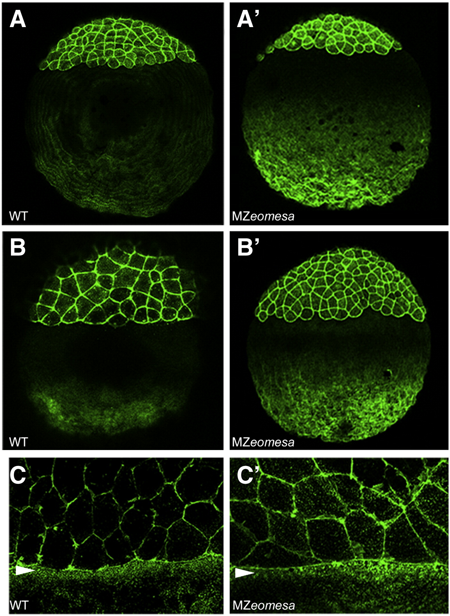
Fig. 6 The actin cytoskeleton is normal is MZeomesa embryos. Confocal projections of lateral views of phalloidin stained embryos. (A-C) wild type and (A′-C′) MZeomesa embryos. (A, A′) sphere stage (B, B′) dome stage, (C, C′) close up of marginal region at 75% epiboly, arrowheads indicate actin band in the YSL.
Figure Data
Acknowledgments
This image is the copyrighted work of the attributed author or publisher, and
ZFIN has permission only to display this image to its users.
Additional permissions should be obtained from the applicable author or publisher of the image.
Reprinted from Developmental Biology, 362(1), Du, S., Draper, B.W., Mione, M., Moens, C.B., and Bruce, A.E., Differential regulation of epiboly initiation and progression by zebrafish Eomesodermin A, 11-23, Copyright (2012) with permission from Elsevier. Full text @ Dev. Biol.