
Image
|
Figure Caption
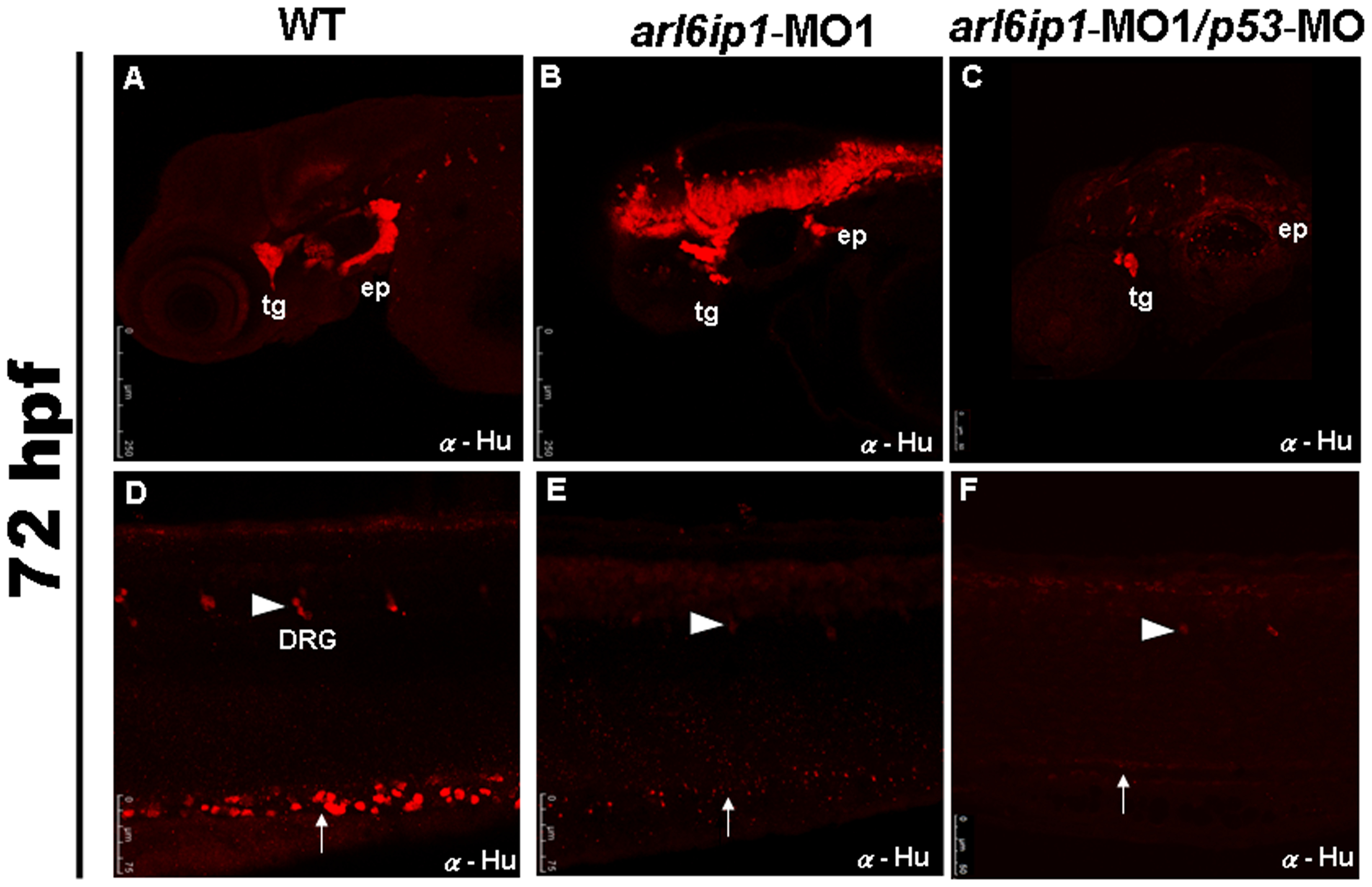
Fig. 3 Neural crest derivatives are reduced in arl6ip1-deficient embryos.
Lateral views (A-F) of 72 hpf embryos processed for anti-Hu immunofluorescence assaying (IFA) to reveal the derivatives of cranial neural crest cells by confocal micrographs. In control embryos (A, D), anti-Hu IFA-labeled trigeminal ganglia (tg), epibranchial ganglia (ep), dorsal root ganglia (drg) (arrowhead), enteric neurons (arrow) all appeared to be reduced either in arl6ip1 knockdown embryos (B, E) or arl6ip1-MO1/p53-MO-injected embryos (C, F).
Figure Data
Acknowledgments
This image is the copyrighted work of the attributed author or publisher, and
ZFIN has permission only to display this image to its users.
Additional permissions should be obtained from the applicable author or publisher of the image.
Full text @ PLoS One