
|
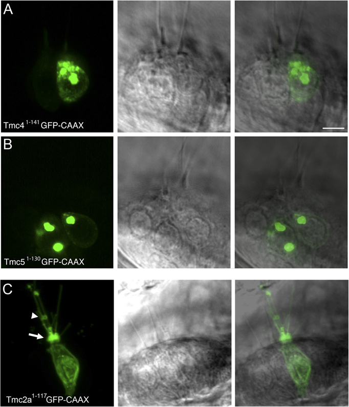
Fig. S7 N-terminal fragments of distantly related Tmcs accumulate within the cell body of hair cells. (A–C) Live images of ampullary hair cells. (A) Tmc4 (1–141 aa) and (B) Tmc5 (1–130 aa) were tagged with GFP-CAAX and mosaically expressed in hair cells using the myo6b promoter. In both low and high expressing hair cells, GFP signal is associated either with intracellular membrane compartments or the basolateral membrane and mostly excluded from hair bundles. Some faint bundle label is seen in the cell of A (n e 24 hair cells for each fragment). (C) Example of morphological defects in a hair cell expressing high levels of Tmc2a1–117-GFP-CAAX. The hair bundle appears amorphous (arrow), and blebbing of the plasma membrane (arrowhead) and unusually long apical processes are evident. In contrast to Tmc4 and Tmc5, large brightly labeled internal compartments were not observed. Cells with morphological defects were excluded from our immunohistochemical and physiological experiments. (Scale bar: 5 μm.)