
|
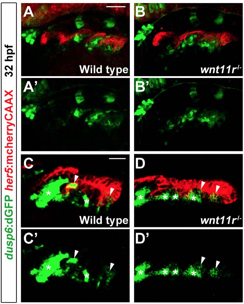
Fig. S6
Fgf activity in the endoderm of wnt11r mutants
(A and B) Confocal projections show Fgf activity as marked by dusp6:dGFP (green) relative to the her5:mCherryCAAX+ pouch endoderm (red) at 32 hpf. In 8/8 wnt11r mutants, dusp6:GFP expression is grossly unaffected compared to wildtype siblings (n=34). Scale bar represents 40 μM. (C and D) Higher magnification and gain-enhanced sections focused on the posterior pouches seen in (A) and (B) show dusp6:dGFP fluorescence within alternating clusters of endodermal epithelial cells (arrowheads), as well as in clusters of adjacent mesodermal cells (asterisks). While pouch outpocketing is delayed in wnt11r mutants, segmental dusp6:dGFP fluorescence is still detectable within both the mesoderm and endoderm. Scale bar represents 20 μM.