
|
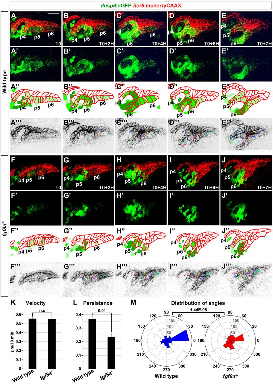
Fig. 5
Requirement for Fgf8a in the directional persistence of pouch cells. (A-J) Representative confocal sections from time-lapse recordings show the development of pouches p4-p6 in a wild type (n=5) and fgf8a mutant (n=3) (see supplementary material Movie 3A,B). her5:mCherryCAAX labels endodermal cell membranes (red) and dusp6:dGFP shows dynamic Fgf activity (green). T0 is 26 hpf. Merged images are shown in A-J and dusp6:dGFP alone in A′′-J′′. Schematics in A′-J′ show the graded intensity of dusp6:dGFP (green) relative to endodermal cells (red). Individual cell tracks used for the quantification are shown in A′′′-J′′′. (K-M) The velocity, persistence of migration and distribution of angles of tracked cells over a 7-h period. For distribution of angles, each bin represents the number of cells moving in a particular direction relative to the last cell position. We tracked the cells of two embryos for each wild type and fgf8a mutant. Mean±s.e.m. and P values are shown. n.s., not significant. Scale bar: 20μm (A-J).