
|
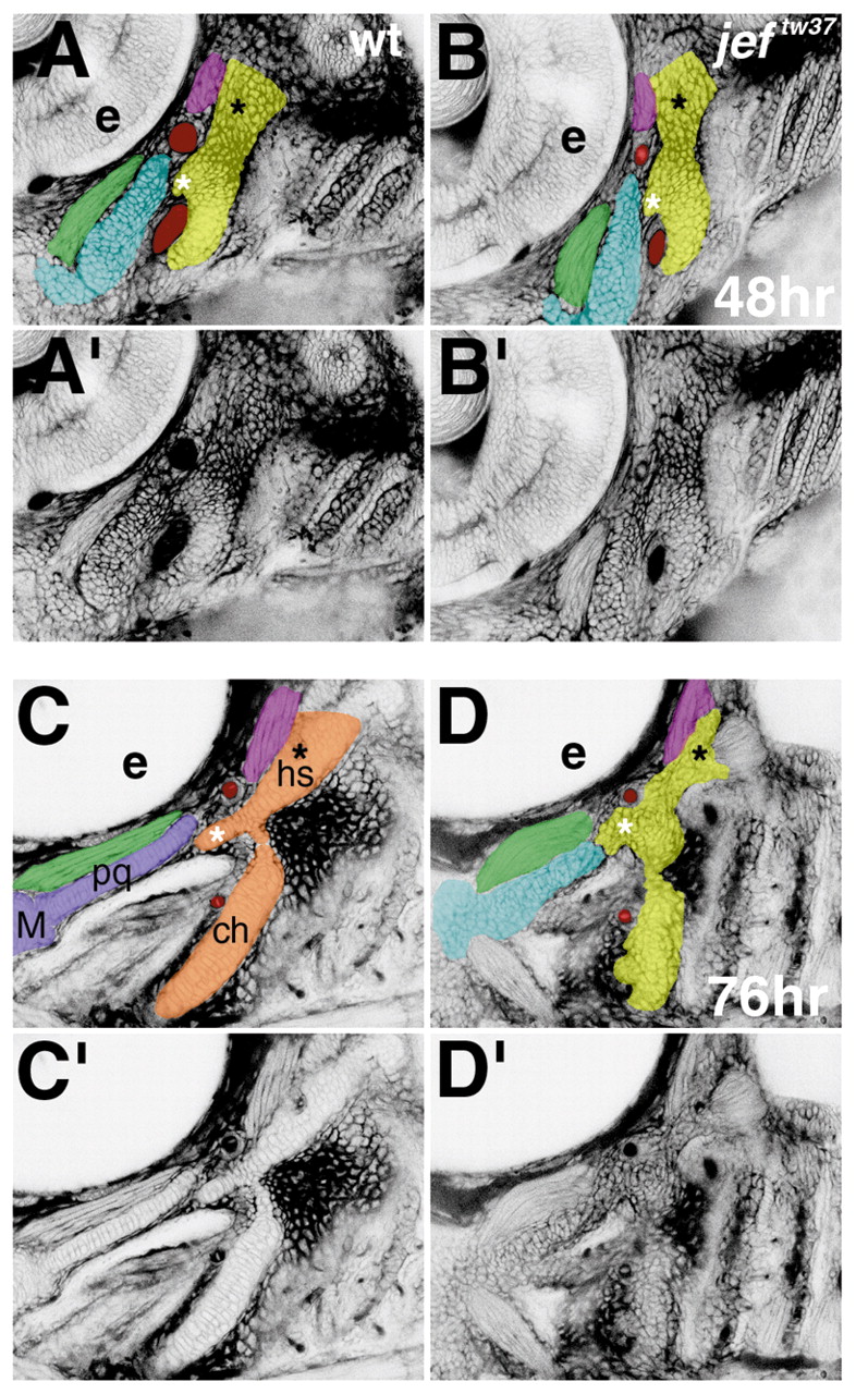
Fig. 8 Prechondrogenic condensations form in jef (sox9a) mutants, but differentiation and morphogenesis fail to occur. Confocal micrographs (lateral views shown as negative images, anterior towards the left) of live wild-type (A,A′,C,C′) and jeftw37 mutants (B,B′,D,D′), stained with BODIPY-ceramide. Cells appear white and interstitial space appears black. (The pharyngeal cavity does not retain the dye, so it also appears white in C-D′). At 48 hours, in wild types (A,A′) and jef mutants for both alleles (B,B′, and data not shown), prechondrogenic condensations have formed. The original images (A′,B′) are pseudo-colored (A,B) to highlight the first (blue) and second (yellow) pharyngeal arch precartilage condensations. Red, first aortic arch; green, adductor mandibulae muscle; pink, constrictor dorsalis premyogenic condensation. At this stage, a contiguous condensation prefigures the dorsal and ventral pharyngeal cartilages, as seen best for the second arch in this focal plane (A-B′). In the second arch precartilage condensation, the symplectic rudiment (white asterisk) and hyomandibular foramen (black asterisk) are apparent in wild types and jef mutants (A-B′). By 76 hours, individuated and precisely shaped dorsal and ventral first and second pharyngeal arch cartilages with highly ordered stacks of chondrocytes have formed in wild types (C,C¢). Original images (C′,D′) are pseudocolored (C,D as in A,B) with overtly differentiated cartilage in wild types colored purple in the first arch and orange in the second arch cartilages. Stacking, individuation, and proper shaping do not occur in jef mutants (D,D′). The precise boundary of the first and second arch is not visible in jef mutants and is approximated in the pseudocolored panel (D). For jeftw37, five mutants and 15 phenotypically wild-type siblings were examined; for jefhi1134, nine mutants and 21 phenotypically wild-type siblings were examined. ch, ceratohyal; e, eye; hs, hyosymplectic; M, Meckel’s; pq, palatoquadrate (see Fig. 1).