
|
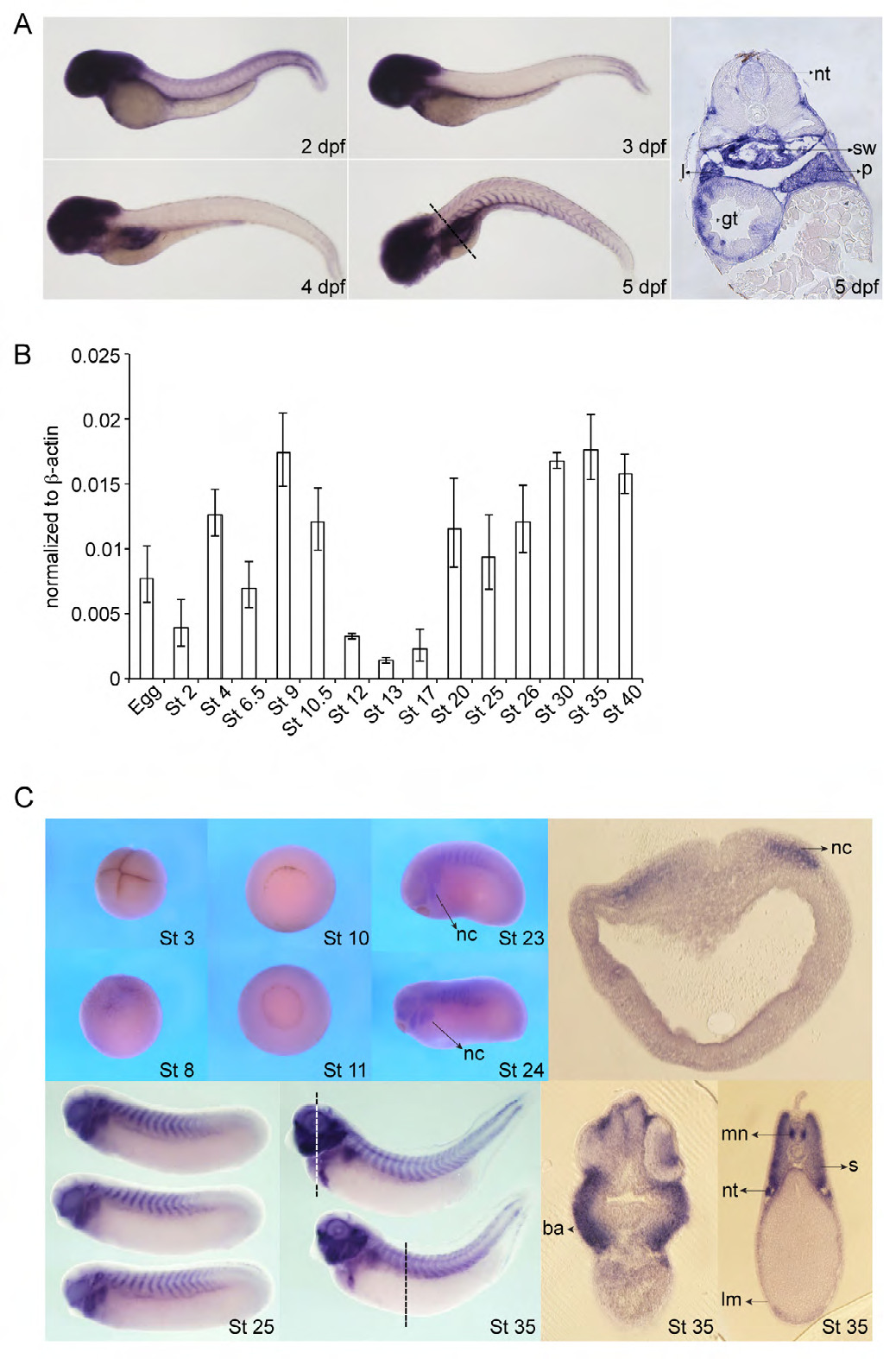
Fig. S1
Later expression of zebrafish mcc and cloning and expression of Xenopus laevis Mcc. (A) Zebrafish mcc expression from 2 to 5 days post fertilization (dpf) by whole-mount in situ hybridization (WISH). mcc transcript levels in the somites decrease and then increase, but persist in head structures and also emerge in the developing digestive system and swim bladder. Section of 5 dpf embryo (far right panel); plane of section indicated by dashed line. Abbreviations: gt, gut tube; l, liver; p, pancreas; sw, swim bladder; nt, neural tube. (B) Expression analysis of Xenopus laevis Mcc by QPCR. Mcc transcripts are detected both maternally (up to stage (St) 8) and zygotically (St 10 and beyond). (C) Xenopus Mcc expression from St 3 to 35 by WISH and vibratome sections of St 18 and 35 embryos at the indicated planes (white and black dashed lines). St 3 (4-cell stage), animal view; St 8, lateral view; St 10 and 11, vegetal view; St 23 and 24, lateral view. Abbreviations: nc, neural crest; ba, branchial arches; mn, motor neurons; nt, nephric tubules; s, somite; lm, lateral mesoderm.