
|
Fig. S4
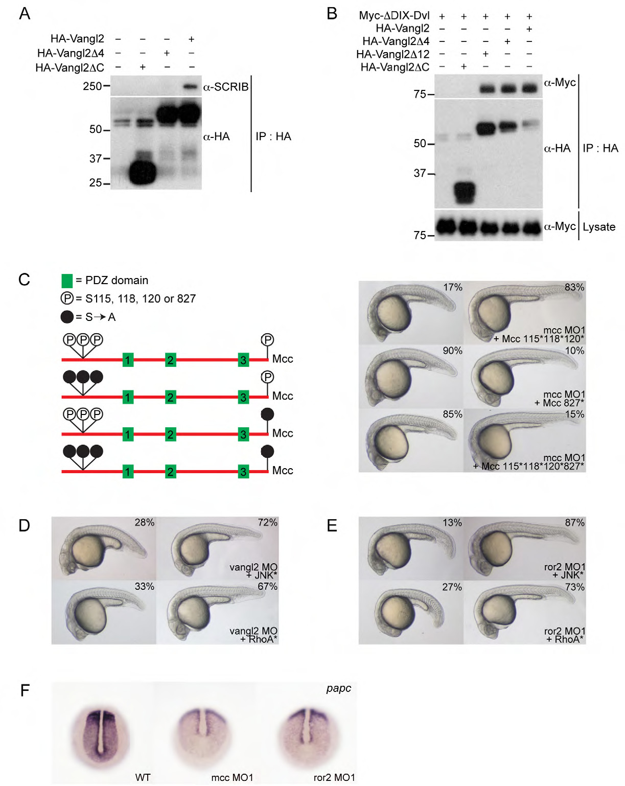
Supporting information for Figure 4. (A) HA-Vangl2 efficiently precipitates endogenous SCRIB in HEK293 cells. Deleting the extreme C-terminal ETSV type I PDZ interaction motif (indicated as Vangl2Δ4) disrupts this interaction. (B) HA-Vangl2, HA-Vangl2Δ4 and HA-Vangl2Δ12 efficiently bind ΔDIX-Dvl—a Xenopus Dishevelled N-terminal truncation that retains its single, central PDZ domain. Deleting the Vangl2 cytoplasmic tail (Vangl2ΔC) eliminates this interaction. These results indicate that Vangl2 binds Dvl via an unconventional PDZ interaction motif within its cytoplasmic tail. (C) Structure of mouse Mcc isoform 2 (not to scale) and location of previously mapped N-terminal (positions 115, 118 and 120) and extreme C-terminal (position 827 (or -1)) serine phosphorylation sites (Pangon et al., 2010; Pangon et al., 2012). Solid black circles indicate mutation of serine residues to non-phosphorylable alanine (S[right arrow]A). Co-injection of 115*, 118* and 120* mutant Mcc mRNA complements mcc loss equally as effectively as wild-type Mcc mRNA (Fig. 2A; Supp. Fig. 2A). In contrast, the C-terminal S827A mutation fails to rescue. (D,E) Activated JNK* (MKK7B2Jnk1a1) and RhoA* (RhoA-G14V) rescue vangl2 and ror2 morphants. (F) Whole-mount in situ hybridization for paraxial protocadherin (papc) expression in the posterior paraxial mesoderm of wild-type and mcc and ror2 morphant embryos at the bud/1-somite stage. Morphants show lower and more diffuse papc expression. (C-E) Morpholino and mRNA concentrations are provided in Supp. Table 3. Phenotypic distributions are indicated as percentages, with scored embryo counts listed in Supp. Table 4.