
|
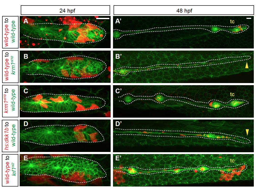
Fig. S9
pLL formation in mosaic embryos that contain krm1nl10, Tg(hsp701:dkk1b-GFP) and lef1nl2 cells (A-E′) Confocal projections of chimeric embryos with rhodamine-labeled donor cells (red) in Tg(cldnB:GFP)-positive hosts. At 24 hpf, donor cells are present in the leading region of primordia in wild-type to wild-type chimeras (A), wild-type to krm1nl10 (B), krm1nl10 to wild-type (C), Tg(hsp701:dkk1b-GFP) to wild-type (D) and wild-type to lef1nl2 mutants (E). At 2 dpf, pLL extension and terminal cluster (tc) formation was seen in wild-type to wild-type (A′), krm1nl10 to wild-type (C’)and wild-type to lef1nl2 (E′). pLL formation was truncated in wild-type to krm1nl10 (B′) and Tg(hsp701:dkk1b-GFP) to wild-type (D′) chimeras. Scale bars=20 μm.