
Image
|
Figure Caption
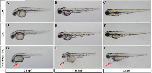
Fig. 2
Overexpression of miR-19b leads to embryonic defects in zebrafish developmental morphology. Representative images of WT (A, B and C), NC (D, E and F), and mir-19b mimic-injected embryos (G, H and I) are shown. MiR-19b-injected embryos displayed severe pericardial edema. All images are at a magnification of 3.2 times. Red arrows: pericardial edema.
Acknowledgments
This image is the copyrighted work of the attributed author or publisher, and
ZFIN has permission only to display this image to its users.
Additional permissions should be obtained from the applicable author or publisher of the image.
Full text @ Cell Physiol. Biochem.