
IMAGE
Fig. S17
- ID
- ZDB-IMAGE-141007-195
- Genes
- Publication
- Bielczyk-Maczyńska et al., 2014 - A loss of function screen of identified genome-wide association study Loci reveals new genes controlling hematopoiesis
- All Figures
- Figures for Bielczyk-Maczyńska et al., 2014
Image
|
Figure Caption
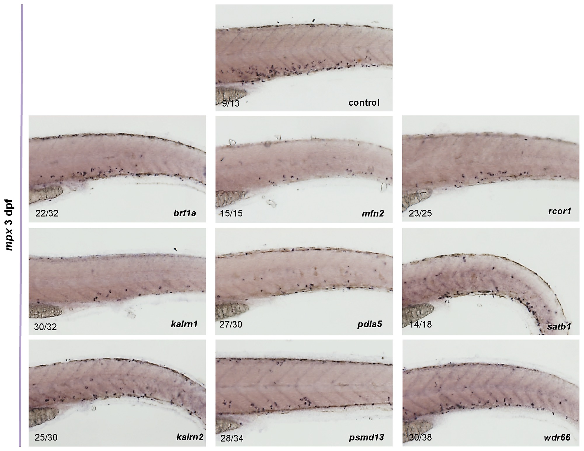
Fig. S17
The expression of mpx in control as well as in brf1a, kalrn1, kalrn2, mfn2, pdia5, psmd13, rcor1, satb1 and wdr66 depleted embryos was assessed by in situ hybridization. For all genes tested the reduced number of mpx positive cells was observed when compared to the control. All embryos are oriented with anterior to the left and dorsal to the top.
Figure Data
Acknowledgments
This image is the copyrighted work of the attributed author or publisher, and
ZFIN has permission only to display this image to its users.
Additional permissions should be obtained from the applicable author or publisher of the image.
Full text @ PLoS Genet.