
|
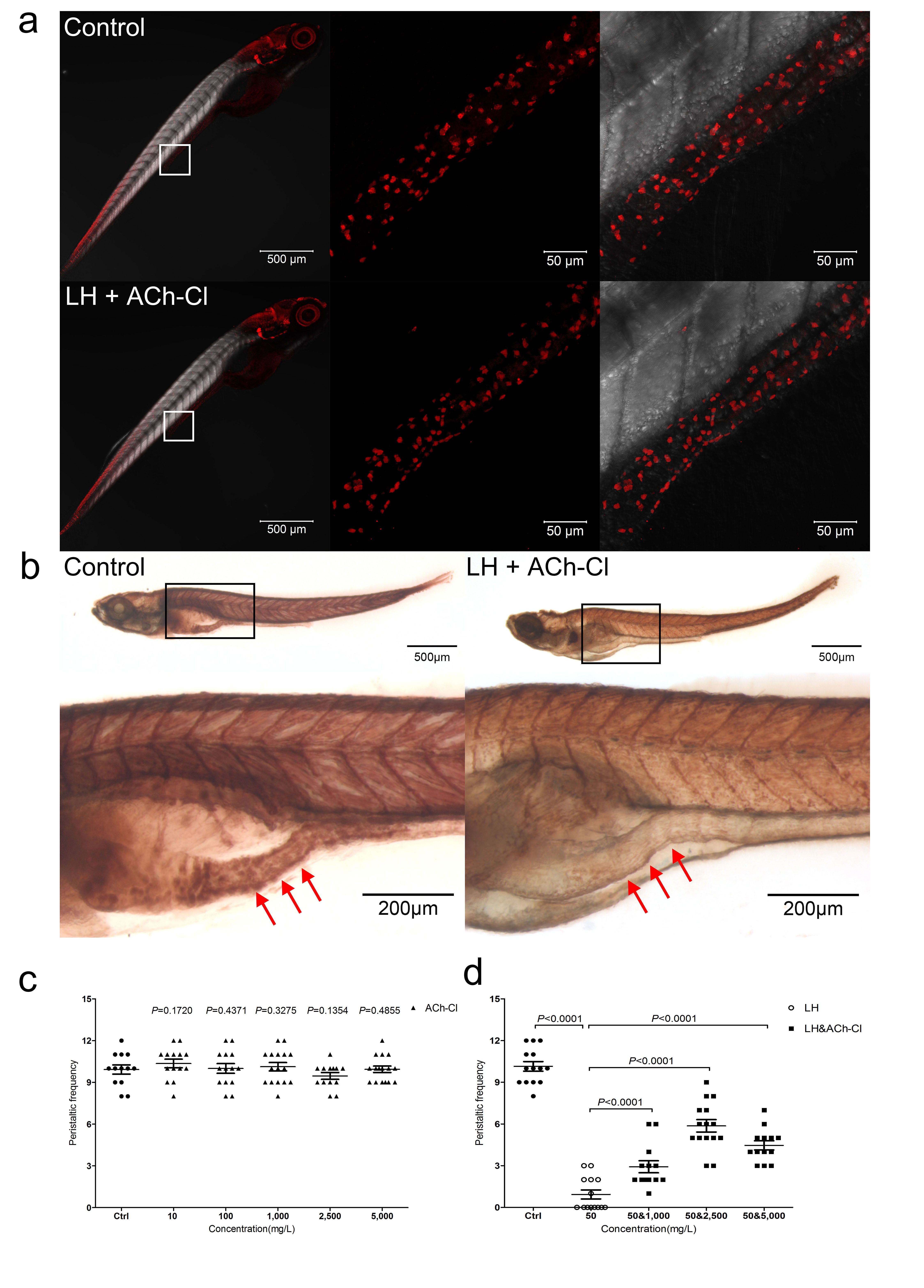
Fig. S4
Effect of combination of ACh-Cl and LH on the gut AChE activity and ENS neuron development.
(a1) Immunohistochemical staining of HuC/D in both control and LH+ACh-Cl treated fish at 6 dpf. The data show no significant difference between the control and chemical application groups. Left pannels show the whole body staining pattern of HuC/D whereas middle and right panels represent high magnifications of the areas that are boxed. Right panels were the images of middle merged with DIC. (b) The AChE activity detection assay reveal that the AChE activity is largely reduced after LH+ACh-Cl treatment compared with control in the gut but not elsewhere in the body. Upper panels show the whole body staining pattern of AChE whereas bottom represent high magnifications of the boxed images accordingly. The red arrows indicate the intestinal bulb region where the peristalsis is identified. (c) Quantification data indicate that there is no obvious promotion effect of exogenous ACh-Cl on gut peristalsis at 6 dpf after incubation for 12 hours with 10, 100, 1000, 2500 and 5000 mg/L. (d) Quantification data show that treatment of exogenous ACh-Cl for 12 hours partially rescue the inhibition phenotype of gut peristalsis caused by LH and the recovery effect is dosage dependent.