
|
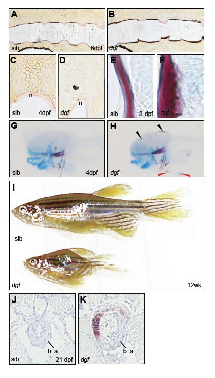
Fig. S1
Van kossa/van Gieson staining shows segmented mineralization of the notochord sheath in siblings (A), in dgf mutants show ectopic calcification of the intervertebral spaces (B). Transverse section through the neuraltube of sibling (C) and mutant with ectopic calcification (D). Alizarin red/Alcian blue stained cleithrum and pectoral fin cartilage of siblings (E) and dgf mutant (D) showing ectopic calcification. Overview of Alizarin red/Alcian blue stained sibling (G) and mutant (H). The black arrowheads indicate cranial calcifications, the red arrowheads point at mineralizations of the skin surrounding the yolk sac and heart (H). dgf mutants can reach adulthood in rare cases but remain smaller (I). Transverse section of alizarin stained juvenile embryos at the bulbus arteriosos (b. a.), no mineralization is visible in silbing (J), circumferential calcification in the dgf mutant (K).