
|
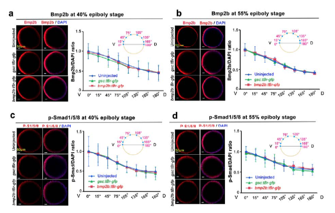
Fig. S5
Bmp2b and p-Smad1/5/8 levels appear unaffected in earlier embryos depleted of organizer Bmp signals. One-cell embryos were injected with 105 pg Tg(-2.067gsc:tBr-gfp) or Tg(-2.272bmp2b:tBr-gfp) plasmid DNA and fixed at 40% and 55% epiboly stages. Uninjected or injected embryos were immunostained using anti-Bmp2b or anti-p-Smad1/5/8 antibody along with DAPI staining. The immunostained embryos were imaged by confocal microscopy from the animal pole. A series of optical equatorial sections at an interval of 3.5 μm were obtained, but only 2 or 3 sections at the blastodermal margin were used for intensity estimation. Eight areas on half of each section (as illustrated above curves), 323.28 μm2 each, were chosen to evaluate immunostaining intensity and DAPI intensity. For each group, 3-5 embryos were analyzed. The ratio of immunostaining intensity to DAPI represented relative immunostaining intensity, and the ratio in the ventralmost position was set to 1.0. (a-d) Representative confocal images and average relative immunostaining intensities. The standard deviations were indicated.