
|
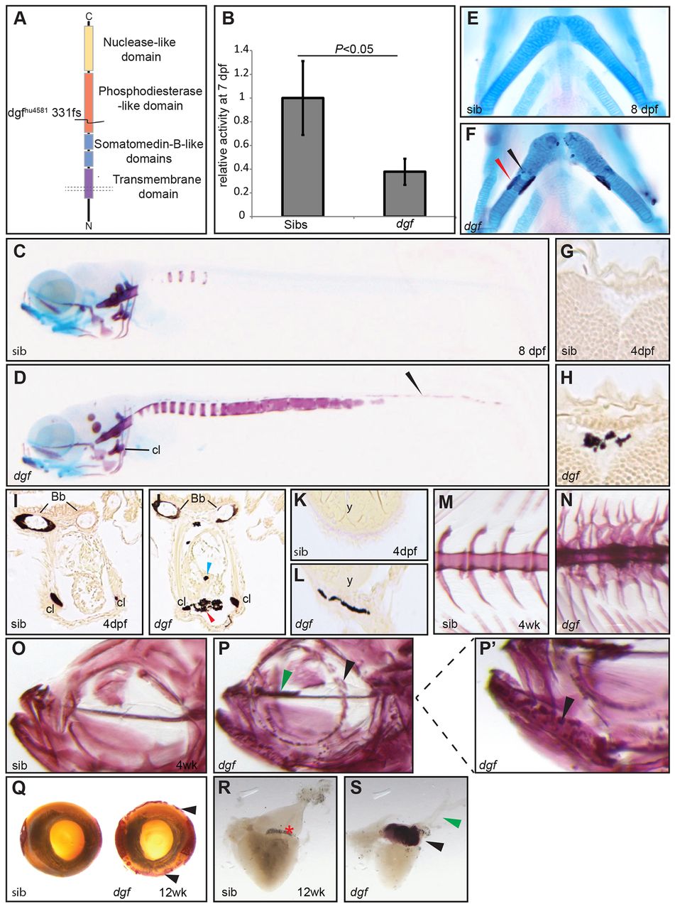
Fig. 1
dgfhu4581 mutants show decreased phosphodiesterase activity and multiple ectopic calcifications. (A) Depiction of the Enpp1 protein structure; the dgfhu4581 allele represents a frame shift at amino acid 331, leading to a premature stop codon. (B) Phosphodiesterase I activity is significantly reduced in the lysate of dgf embryos. Means±1 s.d. are shown. Sib(s), wild-type sibling(s). Alizarin-Red (staining mineralized tissue) and Alcian-Blue (staining cartilage) staining of a sibling embryo (C) and dgf mutant (D) at 8 dpf showing extensive ectopic calcification of the notochord, as well as calcification of the neural tube (D, arrowhead). (E) Ventral view of ceratohyal cartilage element of sibling embryo; in mutant embryos, early onset of perichondral ossification (F, red arrowhead), as well as spots of ectopic cartilage calcification (F, black arrowhead), were observed. van Kossa (brown, staining mineralized tissue) and van Gieson (red, staining osteoid) staining on transverse sections of the brain of a sibling (G) and a dgf mutant with intracranial calcification (H). Transverse section through the heart region of sibling (I) and mutant (J) embryos, both displaying mineralized cleithra (cl) and basobranchial (bb). Mutants (J) in addition display ectopic mineralization between myocard and epicard (red arrowhead) and within the heart (blue arrowhead). (K) Transverse section at the level of the yolk sac of a sibling; (L) the mutant displays ectopic mineralization of the skin. Axial skeleton at the level of the dorsal fin of a sibling (M) and mutant (N) 4-week-old (4wk) fish. Mutants display not only fusion of vertebral bodies but also of neural and haemal arches (N). Alizarin-Red staining of juvenile sibling (O) and mutant (P and enlarged image of the indicated area in P2). Note the ectopic mineralization at the ethmoid plate cartilage element (green arrowhead in P) and nodules of mineralization at the dentary (black arrowheads in P,P2). (Q) Alizarin-Red staining showing ectopic mineralization (black arrowheads) surrounding the eye of a dgf adult mutant (also green arrowhead in P). (R) In the heart of adult zebrafish, no mineralization was visible in siblings. (S) In mutants extensive ectopic calcification was found upon Alizarin-Red staining in the bulbus arteriosus (black arrowhead) but not in the ventral aorta (green arrowhead). Bb, basobranchial; Cl, cleithrum; y, yolk.