
Image
|
Figure Caption
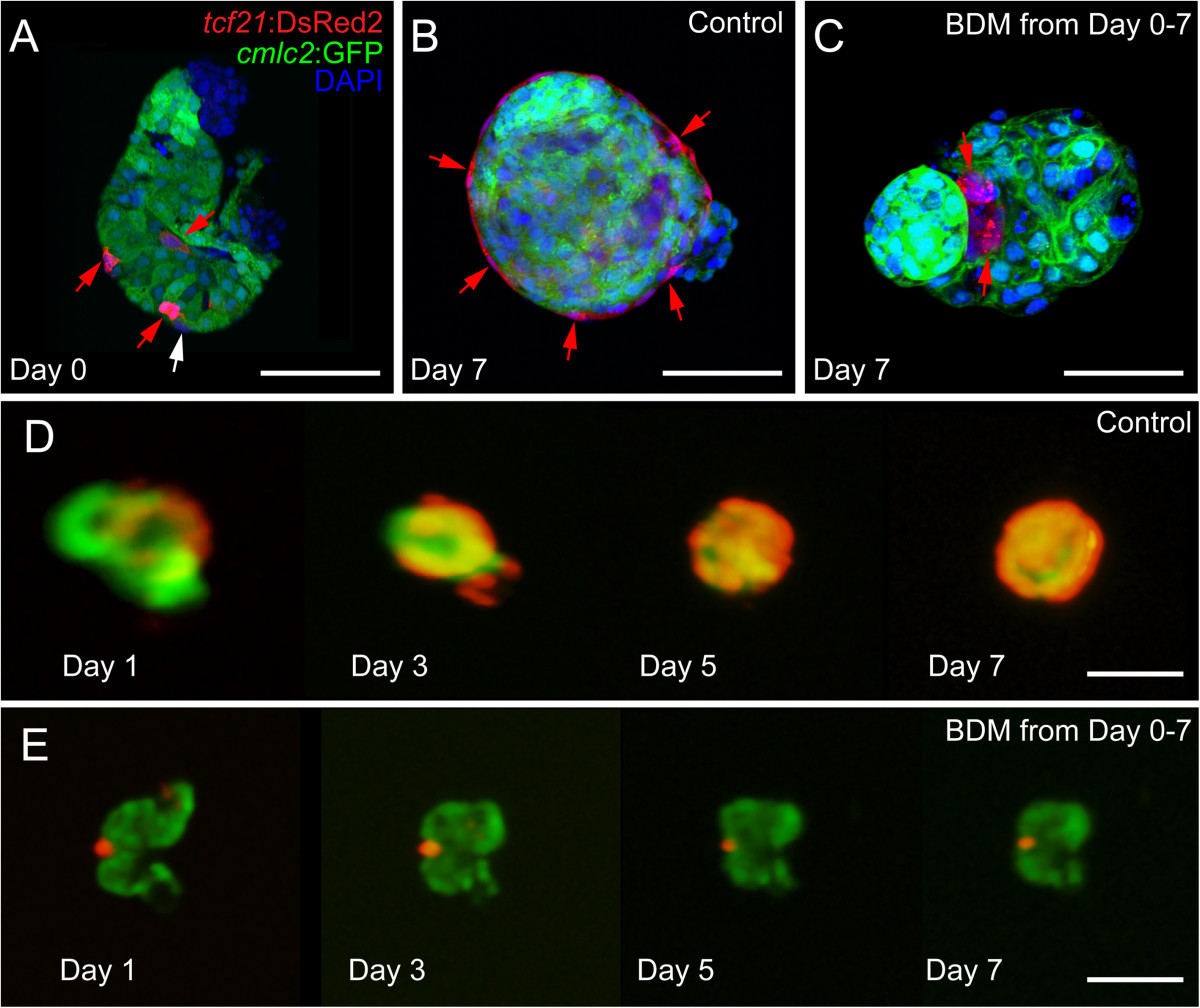
Fig. 8
Inhibiting contraction prevents epicardial development in vitro. Hearts from cmlc2:EGFP; tcf21:DsRed2 larvae were extracted and placed in culture with or without BDM. (A, B, and C) Confocal images of fixed hearts were collected at the time of isolation (A) and after 7 days (B and C) in culture. (B and D) Control hearts (n = 14). (C and E) BDM treated hearts (n = 14). tcf21:DsRed2 marks epicardial cells (red) and cmlc2:EGFP marks cardiomyocytes (green). DAPI (DNA) is in blue in A-C. Scale bars in A-C = 25 microns. Scale bars in D and E = 50 microns.
Acknowledgments
This image is the copyrighted work of the attributed author or publisher, and
ZFIN has permission only to display this image to its users.
Additional permissions should be obtained from the applicable author or publisher of the image.
Full text @ BMC Dev. Biol.