
Image
|
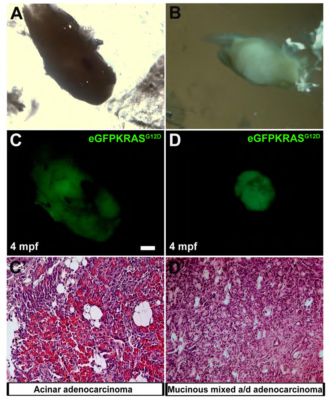
Figure Caption
Fig. S2
Different types of pancreatic cancers. In the panels A and B are reported brightfield images of two different excised pancreatic tumors at 4 months post fertilization. In the panels C and D are reported the relative eGFPKRASG12D positive tumor masses. Two masses corresponded to different types of pancreatic adenocarcinomas: acinar adenocarcinoma with altered structure of exocrine tissue (C′) and mucinous mixed acinar/ductal (a/d) adenocarcinoma (D′). Scale bar is 50 μm.
Acknowledgments
This image is the copyrighted work of the attributed author or publisher, and
ZFIN has permission only to display this image to its users.
Additional permissions should be obtained from the applicable author or publisher of the image.
Full text @ Dis. Model. Mech.