
|
Fig. S6
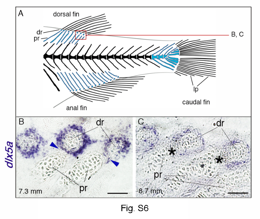
Expression of dlx5a in fin skeletal components of late-stage zebrafish larvae. (A) Overview of the posterior axial skeleton of a one-month-old zebrafish. Endoskeletal fin supports are colored blue and red square indicates the structures analyzed in (B–C). (B, C) Whole mount in situ hybridization for dlx5a on 10 µm parasagittal frozen sections of 7.3 mm and 8.7 mm late-stage zebrafish. As seen in Fig. 8 C in 6.6 mm larvae, dlx5a expression is still detected in cells surrounding the distal radials (dr) in 7.3 mm larvae, including in the zone of segmentation (ZS) (B, blue arrowheads) when the radial segmentation is almost completed. Later, after segmentation (8.7 mm), dlx5a expression is maintained but decreases in cells surrounding the distal radials and is no longer detected in the ZS (black asterisks) (C). dr, distal radials; lp, lepidotrichia; pr, proximal radials. Scale bars B–C 10 μm.