
Image
|
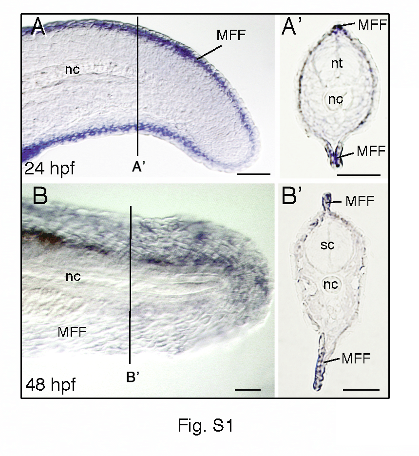
Figure Caption
Fig. S1
Expression patterns of dlx5a during zebrafish median fin development. Whole mount in situ hybridization for dlx5a on lateral view of the posterior axis of 24 hpf (A) and 48 hpf (B) zebrafish embryos and on 10 µm parasagittal frozen sections (A2, B2) at the level indicated in (A, B). At 24 and 48 hpf, dlx5a expression is limited to apical ectodermal cells of the median fin fold (MFF). nc, notochord; nt, neural tube; sc, spinal cord. Scale bars 50 μm.
Acknowledgments
This image is the copyrighted work of the attributed author or publisher, and
ZFIN has permission only to display this image to its users.
Additional permissions should be obtained from the applicable author or publisher of the image.
Full text @ PLoS One