
|
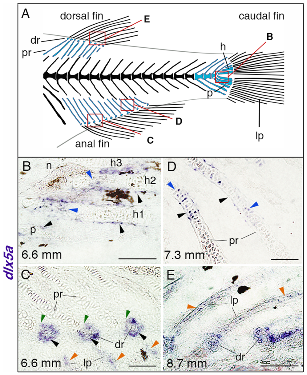
Fig. 8
dlx5a expression during unpaired fin skeletogenesis.
(A) Overview of the posterior axial skeleton of a one-month-old zebrafish. Endoskeletal fin supports are colored in blue and red squares indicate the structures analyzed in (B–E). (B–E) Whole mount in situ hybridization for dlx5a on 10 µm parasagittal frozen sections of late-stage zebrafish from 6.6 mm (16 days post-fertilization) to 8.7 mm (1 month post-fertilization). In 6.6 mm larvae, dlx5a is expressed in the perichondrium of the hypurals 1 to 3 (h1-3) and of the parahypural (p) (B, black arrowheads) as well as in maturing chondrocytes of h1 and h2 (B, blue arrowheads). The transcripts are also detected in cells surrounding the distal radials (dr) during radial segmentation (C, black arrowheads), notably in the zone of segmentation (C, green arrowheads), and in the developing lepidotrichia (lp, C, orange arrowheads). Later, dlx5a expression is observed in the perichondrium (D, black arrowheads) and in maturing chondrocytes (blue arrowheads) of proximal radials (pr) of 7.3 mm larvae. In 8.7 mm larvae, dlx5a expression is maintained in the well-developed lepidotrichia (lp) (E, orange arrowheads). Black-brown patches in B–C, E are melanophores. Scale bars B, E 20 μm, C–D 10 μm.