
|
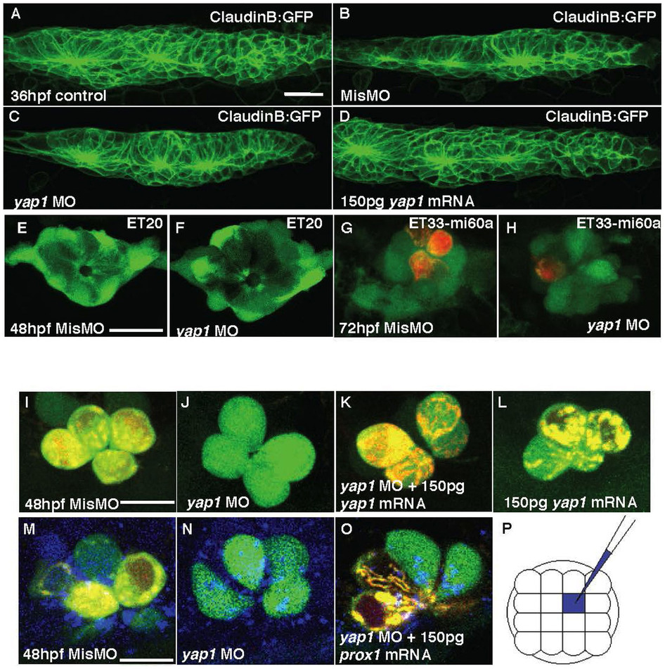
Fig. 4
(A–D) Primordium I (PrimI) in yap1 morphants. (E) Size of PrimI at 36 hpf. (E, F) Mantle cells of neuromast in 72 hpf ET20 yap1 morphants. (G–H) Mechanoreceptors of neuromast (red, DiAsp) in 72 hpf ET33-mi60a embryos, support cells (green). (I–L) hair cells. (J–L) Loss of yap1 abolished the functionality of hair cells in SqET4 transgenics, stained with DiAsp (red); it was rescued with yap1 mRNA injection. (M–O) Mismatch MO, yap1 MO or yap1 MO with prox1 mRNA were co-injected with a marker (Cascade Blue) into single cell of SqET4 embryos at 16-cell stage. (P) The schematic drawing of injected single cell (blue), any one of the middle four cells, at 16-cell stage. Scale bar = 10 μm.