
|
Fig. 8
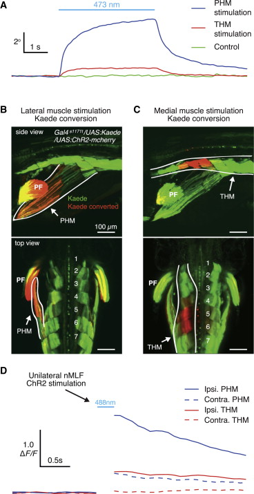
Specific Muscle Activation Generates Tail Deflections
(A) Change in tail angle in Gal4s1171t/UAS:ChR2 fish elicited by ipsilateral hypaxial muscle stimulation with a stationary vertically oriented optic fiber (105 μm fiber diameter, continuous beam 2 mW/mm2). Blue line depicts the epoch of ChR2 stimulation. The blue trace represents the average change in tail angle when lateral muscle regions in the vicinity of myotomes 4–6 were stimulated (n = 6). The red trace represents the average change in tail angle when muscle regions just lateral to the midline were stimulated (n = 6). The green trace represents the average change in tail angle in Gal4s1171t/UAS:GFP control fish (n = 3).
(B) Confocal image projection from the side and top of a Gal4s1171t/UAS:ChR2/UAS:Kaede fish that underwent Kaede conversion at a lateral muscle site, which produced a substantial tail deflection angle. This conversion labeled the posterior hypaxial muscle group (PHM) outlined in white. Numbers indicate myotome.
(C) Confocal image projection from the side and top of a Gal4s1171t/UAS:ChR2/UAS:Kaede fish that underwent Kaede conversion at a medial muscle site, which produced a small, but detectable tail deflection. This conversion labeled the trunk hypaxial muscle group (THM) outlined in white.
(D) Average calcium responses in ipsilateral PHM (blue), ipsilateral THM (red), contralateral PHM (blue dash), and contralateral THM (red dash) following unilateral nMLF optical stimulation in Gal4s1171t/UAS:ChR2/UAS:GCaMP6s fish (n = 5). Blue line depicts a 200 ms epoch of ChR2 stimulation. ChR2 was stimulated using the minimum laser power required to produce a tail deflection. n values indicate number of fish. PF, pectoral fin.
See also Figure S8.