
|
Fig. 7
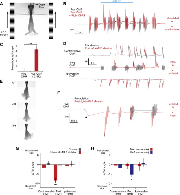
Role of Lateral nMLF Neurons in the Optomotor Response
(A) Schematic of the optomotor assay. Animal is head-embedded in agarose with the tail free. Two LCD screens on each side of the fish display caudal to rostral moving gratings for forward OMR stimulation. A third screen (not shown) is added in front of the animal to create rotating gratings for contraversive and ipsiversive OMR stimulation.
(B) Change in tail angle in a Gal4s1171t/UAS:ChR2 fish elicited by continuous forward OMR stimulation (black) or by both OMR stimulation and ChR2 activation of the right nMLF (red). Blue line depicts the epoch of ChR2 stimulation.
(C) Increase in ipsilateral tail angle during forward OMR swim bouts induced by ChR2 stimulation (n = 21; p < 10-6; t(20) = -5.26).
(D) Change in tail angle in a Gal4s1171t/UAS:GFP fish evoked by three different OMR stimuli before (black) and after (red) ablation of lateral neurons in the left nMLF. All traces are from the same fish.
(E) Projection of video frames displaying tail position of three fish (right ablated, control and left ablated) for an entire forward OMR trial.
(F) Change in tail angle evoked by a forward OMR stimulus before (gray) and after (red) ablation of lateral neurons in the right nMLF. Black arrowhead denotes a left tail deflection that precedes swimming. Traces are from the same fish.
(G) In ablated fish (red bars) swims are biased toward the intact side for forward OMR (n = 31; paired t test, p < 10-6; t(30) = 6.96). Turns evoked by contraversive (n = 28; p = 0.076; t(27) = 1.84) or ipsiversive (n = 28; p = 0.55; t(27) = 0.59) grating stimuli are unchanged. In control fish (gray bars), no biases were observed for forward (n = 10; p = 0.70; t(9) = 0.39), contraversive (n = 8; p = 0.52; t(7) = -0.67), or ipsiversive (n = 8; p = 0.86; t(7) = -0.17) stimuli.
(H) Unilateral superficial small neuron ablations (blue bars) generated a bias in OMR forward swims toward the intact side (n = 13; paired t test, p < 0.02; t(12) = 2.82). Responses to contraversive (n = 11; p = 0.63; t(10) = 0.48) or ipsiversive (n = 11; p = 0.88; t(10) = -0.14) stimuli were unchanged. Unilateral MeL neuron ablations (red bars) did not affect forward swims (n = 8; p = 0.62; t(7) = 0.51), or turns to contraversive (n = 8; p = 0.97; t(7) = 0.03) or ipsiversive (n = 7; p = 0.63; t(6) = 0.51) stimuli. n values indicate number of fish. Error bars indicate SEM.
See also Figure S7 and Movies S4 and S5.