
Image
|
Figure Caption
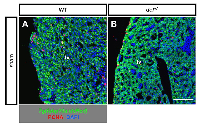
Fig. S4
Comparison of cell proliferation in the adult liver in the wild-type and def+/- sham controls. Representative images of immunostaining of PCNA (in red) in the Tg(fabp10a:RFP) background, in which hepatocytes are genetically labelled by expressing the red fluorescent protein (in green), showed that proliferating cells were rarely detected along the epithelial edge of an adult liver in either the wild-type or def+/- mutant sham controls. Nuclei were stained with DAPI (blue). lv, liver tissue. Scale bar: 75 μm.
Acknowledgments
This image is the copyrighted work of the attributed author or publisher, and
ZFIN has permission only to display this image to its users.
Additional permissions should be obtained from the applicable author or publisher of the image.
Full text @ PLoS One