
Image
|
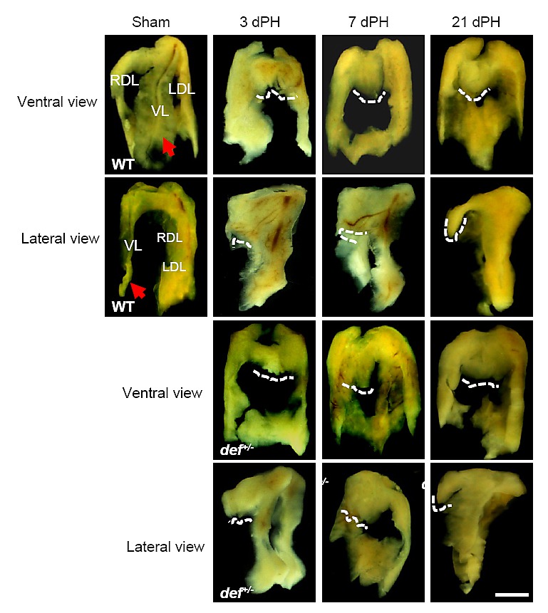
Figure Caption
Fig. S2
The def+/- mutant showed defective lobe structure recovery at the amputation site after PH. Lower magnifications of the ventral and lateral views of the gross morphology of the liver 3, 7 and 21 days after PH. The images for 7 days after PH correspond to the closer view of the amputation site shown in Figure 1d. The white, dashed line outlines the amputation site on the ventral tip. The red arrow highlights the ventral lobe in the sham control. VL: ventral lobe; LDL: left dorsal lobe; RDL: right dorsal lobe. Scale bar: 1 cm.
Acknowledgments
This image is the copyrighted work of the attributed author or publisher, and
ZFIN has permission only to display this image to its users.
Additional permissions should be obtained from the applicable author or publisher of the image.
Full text @ PLoS One