
|
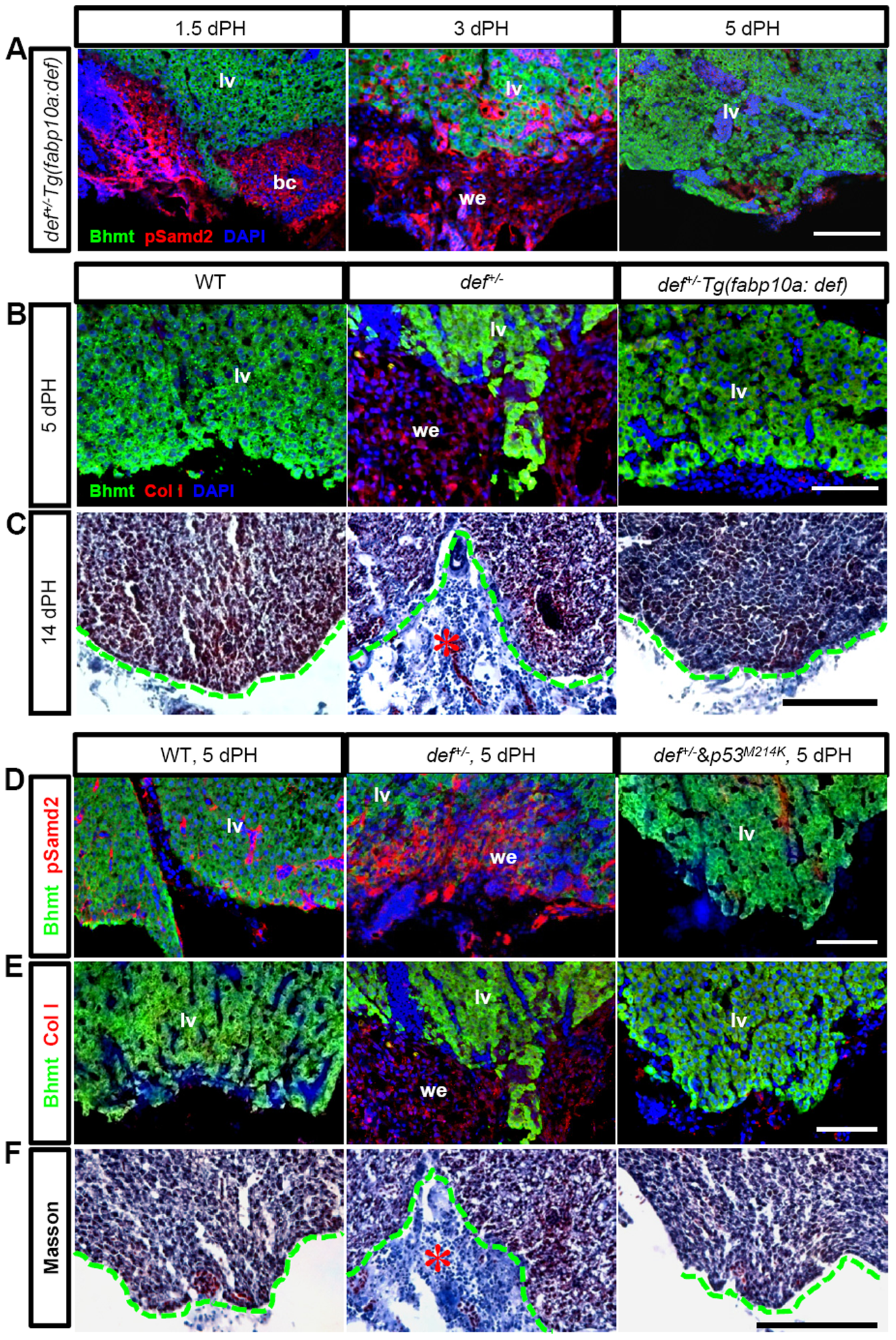
Fig. 10
Formation of the fibrotic scar in def+/- fish after PH was mediated by the activation of the p53-pathway.
(A) Immunostaining of pSmad2 (red) and Bhmt (green) in the wound dermis in def+/-Tg(fabp10:def)+/- progeny 1.5, 3 and 5 days after PH, respectively. (B) Immunostaining of ColI (red) and Bhmt (green) in the wild-type, def+/- and def+/-Tg(fabp10:def)+/- progeny 5 days after PH. (C) Masson staining of the amputation site (indicated by a green dashed line) in the wild-type, def+/- and def+/- Tg(fabp10:def)+/- progeny 14 days after PH. (D,E) Immunostaining of pSmad2 (red) and Bhmt (green) (D) and of ColI (red) and Bhmt (green) (E) in the wound epidermis in the wild-type, def+/- and def+/-p53M214K/M214K 5 days after PH. (F) Masson staining of the amputation site in the wild-type, def+/- and def+/-p53M214K/M214K progeny 14 days after PH. Fibrotic tissue in the def+/- fish is highlighted by a red asterisk. In (A,B,D,E), nuclei were stained with DAPI (blue). In each case, more than 10 sections from at least three wild-type or def+/- mutant fish were examined. Representative images are shown here. bc, blood clot; lv, liver tissue; we, wound epidermis. Scale bars: 150 μm (C,F) and 75 μm (A,B,D,E).