IMAGE

Fig. 3

ID
ZDB-IMAGE-140808-3
Source
Figures for Monteiro et al., 2014
Image
Figure Caption

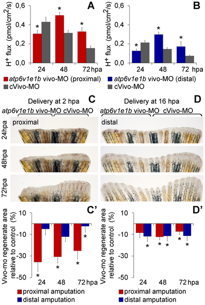
Fig. 3 Vivo-morpholino mediated atp6v1e1b knockdown impairs regeneration and decreases H+ flux.

(A, B) H+ flux during regeneration after vivo-MO mediated atp6v1e1b knockdown, 2 h after proximal (A) and distal (B) amputation. *statistical significant results (p<0.05). (C, D) Caudal fin regeneration after atp6v1e1b vivo-MO delivery to atp6v1e1bhi577aTg/+ (AB) fish, 2 h after proximal amputation (C) and 16 h after distal amputation (D). (C′, D′) Percentage of regenerate area in atp6v1e1b knocked down regions compared to the control vivo-MO (cVivo-MO). Vivo-MOs were delivered 2 h (C′) or 16 h (D′) after both proximal and distal amputation. hpa: hours post amputation. For each panel, n = 8.

Figure Data
Acknowledgments
This image is the copyrighted work of the attributed author or publisher, and ZFIN has permission only to display this image to its users. Additional permissions should be obtained from the applicable author or publisher of the image. Full text @ PLoS One