
|
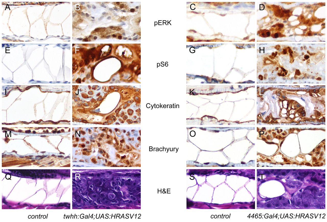
Fig. 2 Immunohistochemical features of the zebrafish notochord tumors. 7-dpf larvae were examined by immunohistochemical techniques. The left two columns show control and twhh:Gal4;UAS:HRASV12, and the right two columns show control and 4465:Gal4;UAS:HRASV12. (A–D) pERK staining in control embryos (A,C) showed minimal nuclear positivity in the notochord cells. EGFP-HRASV12 embryos showed focal, strong nuclear and weak cytoplasmic positivity in tumor cells (B,D). (E–H) pS6 staining in control embryos was negative in the notochord (E,G), but the tumor cells showed strong nuclear and weak cytoplasmic positivity. (I–L) Cytokeratin staining showed cytoplasmic positivity in normal notochord cells (I,K) and in the tumor (J,L). (M–P) Brachyury staining demonstrated nuclear and, to a lesser extent, cytoplasmic positivity in normal notochord cells (M,O) and in the tumor (N,P). (Q–T) Corresponding histology of normal notochord (Q,S) and the notochord tumor (R,T).