
|
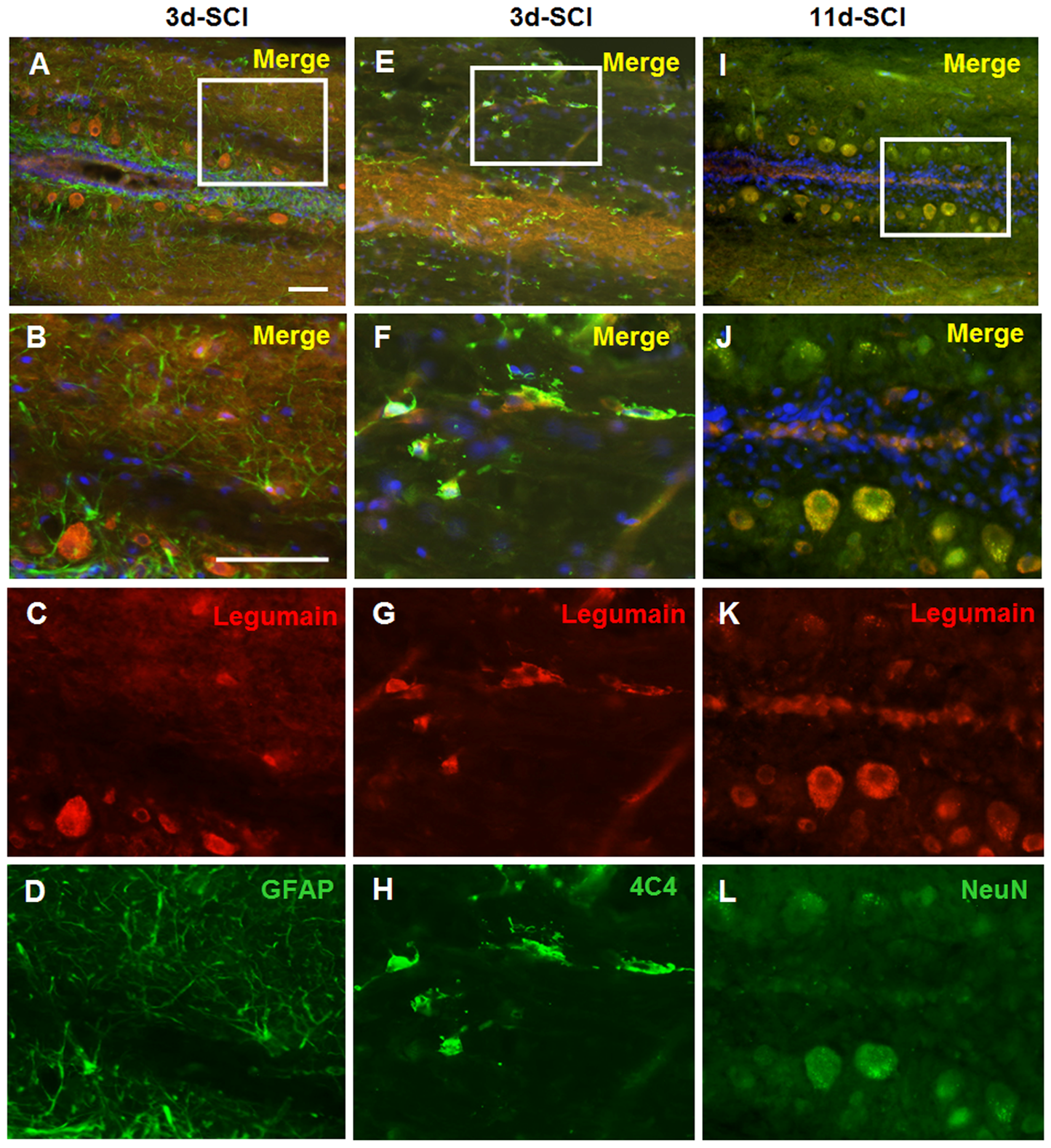
Fig. 4
Neurons and macrophages/microglia express Legumain in the caudal spinal cord after SCI.
Spinal cord sections from fish 3 days after SCI were used for double staining of Legumain and GFAP (A–D) or 4C4 (E–H). Spinal cord sections from fish 11 days after SCI were used for double staining of Legumain and NeuN (I–L). B, F, and J are the magnifications of A, E and I, respectively. No co-localization of Legumain and GFAP is observed (A–D). Double immunostaining of Legumain with 4C4 antibody identifies the positive small cells as macrophages/microglia (E–H). Double staining with NeuN shows that neurons express Legumain at 11 days after SCI (I–L). n = 3 experiments. Scale bar, 50 μm.