
|
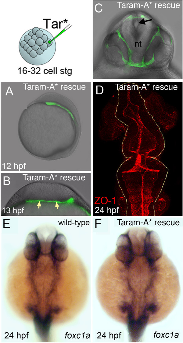
Fig. 5
Mesoderm rescues neural tube morphogenesis in MZoep embryos. (A) Lateral view of an MZoep embryo previously injected with the activated form of the TGF-β receptor Taram-A* (Tar*) into a single blastomere. Rescued head mesoderm expresses GFP (green). (B) Transverse section of Taram-A*-injected embryos show rescued mesoderm (green and arrowed) underlying neural plate at 13 hpf. (C) By 24 hpf rescued mesoderm almost surrounds the neural tube (nt) in MZoep embryos. The morphology of the neural tube is rescued and contains a single well-defined midline lumen (black arrow). (D) Rescued neural lumen morphology revealed by ZO-1 expression. (E,F) Identity, distribution and presence of rescued mesoderm, confirmed by the mesendoderm marker foxc1a, in wild-type and Taram-A*-injected MZoep embryos. GFP, green fluorescent protein; hpf, hours post fertilization; MZoep, maternal-zygotic one-eyed pinhead; TGF-β, transforming growth factor beta; ZO-1, zonula occludens 1.