
|
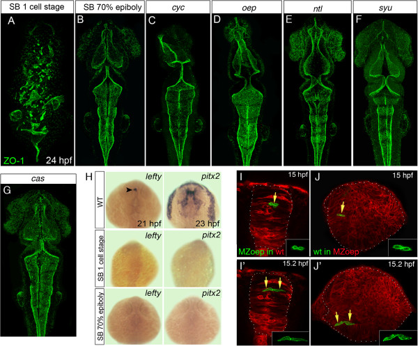
Fig. 4 Loss of mesoderm, but not Nodal signaling, leads to neural tube defects. (A,B,C,D,E,F,G) Projection of confocal z-series showing dorsal view of 24 hpf zebrafish embryos staining for the apical marker ZO-1 (green). (A) Disrupted ventricular organization of embryo treated with the Nodal inhibitor SB-431542 from the one-cell stage. (B) Treatment with SB-431542 from 70% epiboly does not cause neural tube defects. (C,D) Ventricle organization in Nodal mutants cyc and oep is largely normal. (E,F) Ventricle organization in ntl and syu mutants is normal. (G) Ventricle organization in the endoderm mutant cas mutants is normal. (H) Expression of the Nodal-dependent markers lefty and pitx2 show the SB-431542 drug is an efficient blocker of Nodal signaling. All embryos between 21 and 23 hpf, dorsal view. Arrowhead in (H) indicates asymmetric lefty expression at 21 hpf. (I,I′) Two frames from time-lapse sequence show transplanted MZoep cells (green) integrate and divide to make mirror-image daughters just like host cells in a wild-type embryo at 15 hpf. (J,J′) Two frames at 15 hpf from time-lapse sequence show wild-type cells (green) divide to make mirror-image daughters after transplantation to a MZoep embryo. Unlike divisions in wild-type embryos, however, these divisions can be far from their normal location at the midline. hpf, hours post fertilization; MZoep, maternal-zygotic one-eyed pinhead; wt, wild-type; ZO-1, zonula occludens 1.