
|
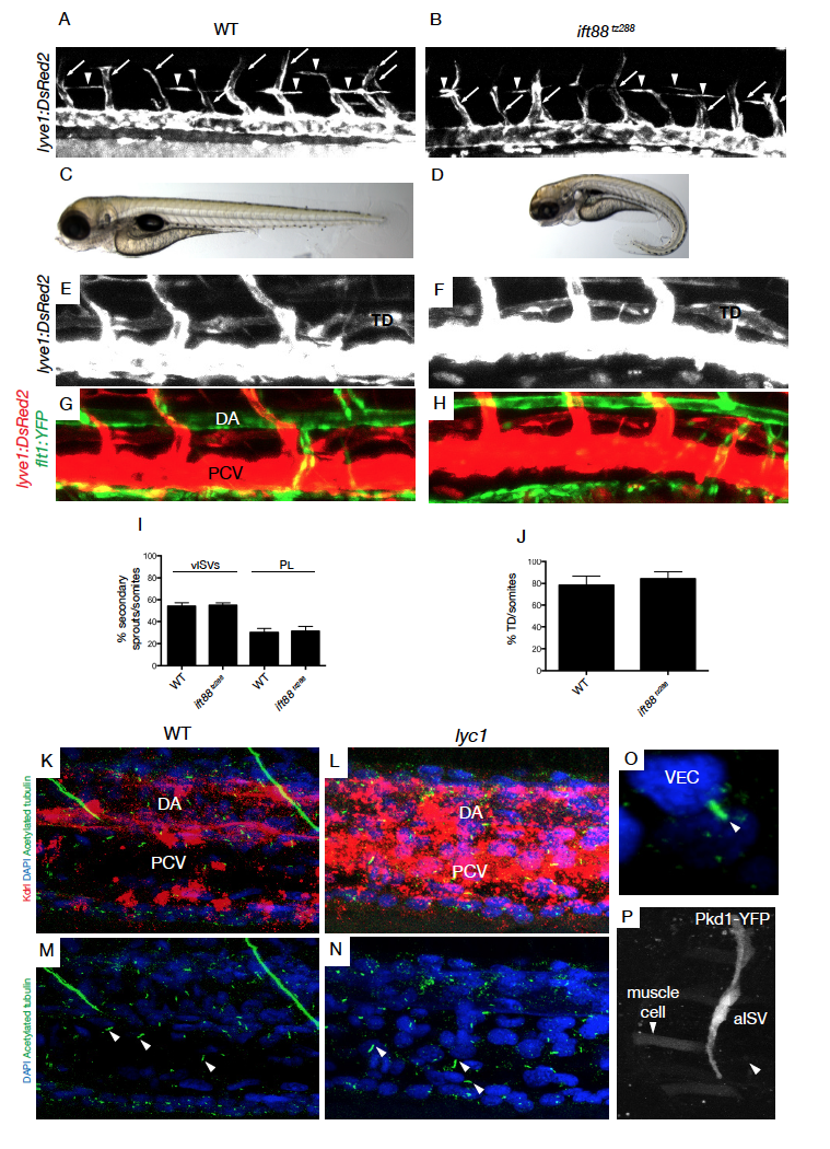
Fig. S8
No evidence for a contribution of primary cilia to lymphangiogenesis. Related to Discussion section.
(A-B) The vasculature Tg(lyve1:DsRed2)nz101
in (A) wild-type sibling and (B) ift88tz288 mutant embryos at 56 hpf (arrowheads indicate parachordal lymphangioblasts and white arrows indicate venous sprouts).(C-D) Overall morphology of (C) wild-type siblings and (D) ift88tz288 mutants at 5 dpf. (E-H) The vasculature Tg(lyve1:DsRed2nz101; flt1:YFPhu4624Tg) of (E,G) WT and (F,H) ift88tz288 mutants at 5 dpf.
(I-J) Quantification of (I) secondary sprouts in WT (n=12) and ift88tz288 (n=10) at 56 hpf and (J) thoracic duct extent in WT (n=16) and ift88tz288 (n=18).
(K-N) Overview of primary cilia localization in the trunk of (K,M) WT (n=4) and (L,N) lyc1 mutants (n=3) embryos at 30 hpf, stained for blood vessels (Kdrl-Cherry), nuclei (DAPI) and primary cilia (Acetylatedtubulin) markers. Arrowheads indicate example of discrete primary cilia. (O) An individual representative primary cilium in a lyc1 embryo.
(P) Transient expression of a Pkd1-YFP BAC construct in an arterial intersegmental vessel and adjacent muscle cells at 4 dpf. DA: Dorsal Aorta, PCV: Posterior Cardinal Vein, TD: Thoracic duct