
Fig. 1
|
Fig. 1
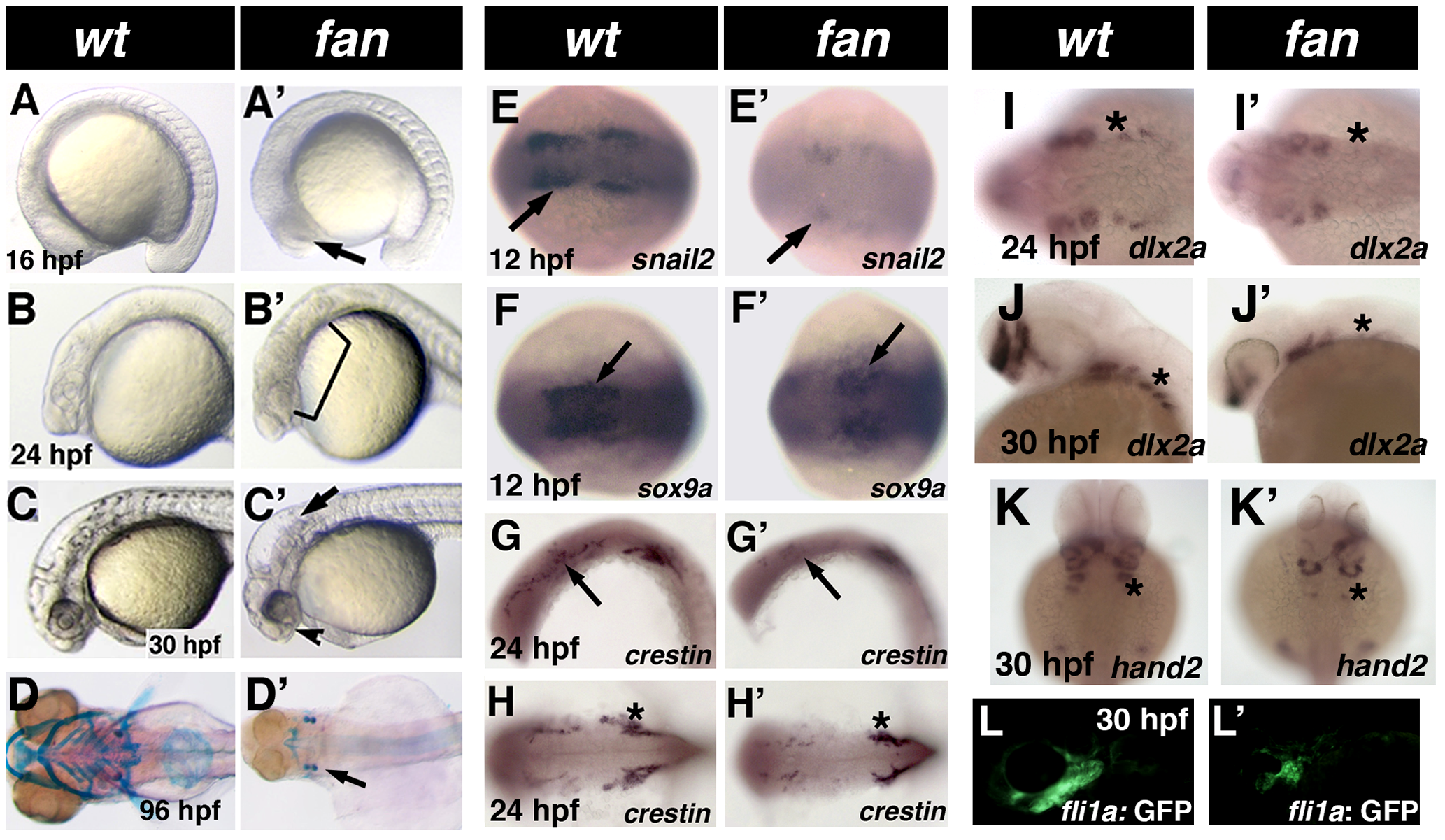
Phenotype of fan mutants.
(A–C2) Live images of developmentally staged wild type (A–C) and fan mutant (A2–C2) zebrafish. Arrow in A2 indicates necrotic cells in the presumptive eye region. Bracket in B2 shows necrosis in neural and pharyngeal arch tissues. Arrow in C2 points to the distinct hydrocephaly in fan mutant hind brain ventricles, arrowhead indicates incomplete choroid fissure closure and craniofacial defects. (D–D2) Alcian blue stained pharyngeal arch cartilages in 4dpf wild type (D) and fan mutant (D2). (E–K2) Whole mount ISH images of wild type (E–K) and fan mutants (E2–K2) for neural crest markers at indicated developmental stage. Arrows in (E2–G2) and asterisks in (I2–K2) indicate reduced gene expression in fan mutant embryos. Interestingly, fan mutant embryos exhibit similar crestin expression in the trunk NCCs (asterisks in H, H2) but reduced expression in cranial NCCs. (L–L2) Tg(fli1a:EGFP)/fan mutants (L2) exhibit reduced GFP expression in the pharyngeal arch region as compared to wild type embryos (L).