
Image
|
Figure Caption
Fig. 3
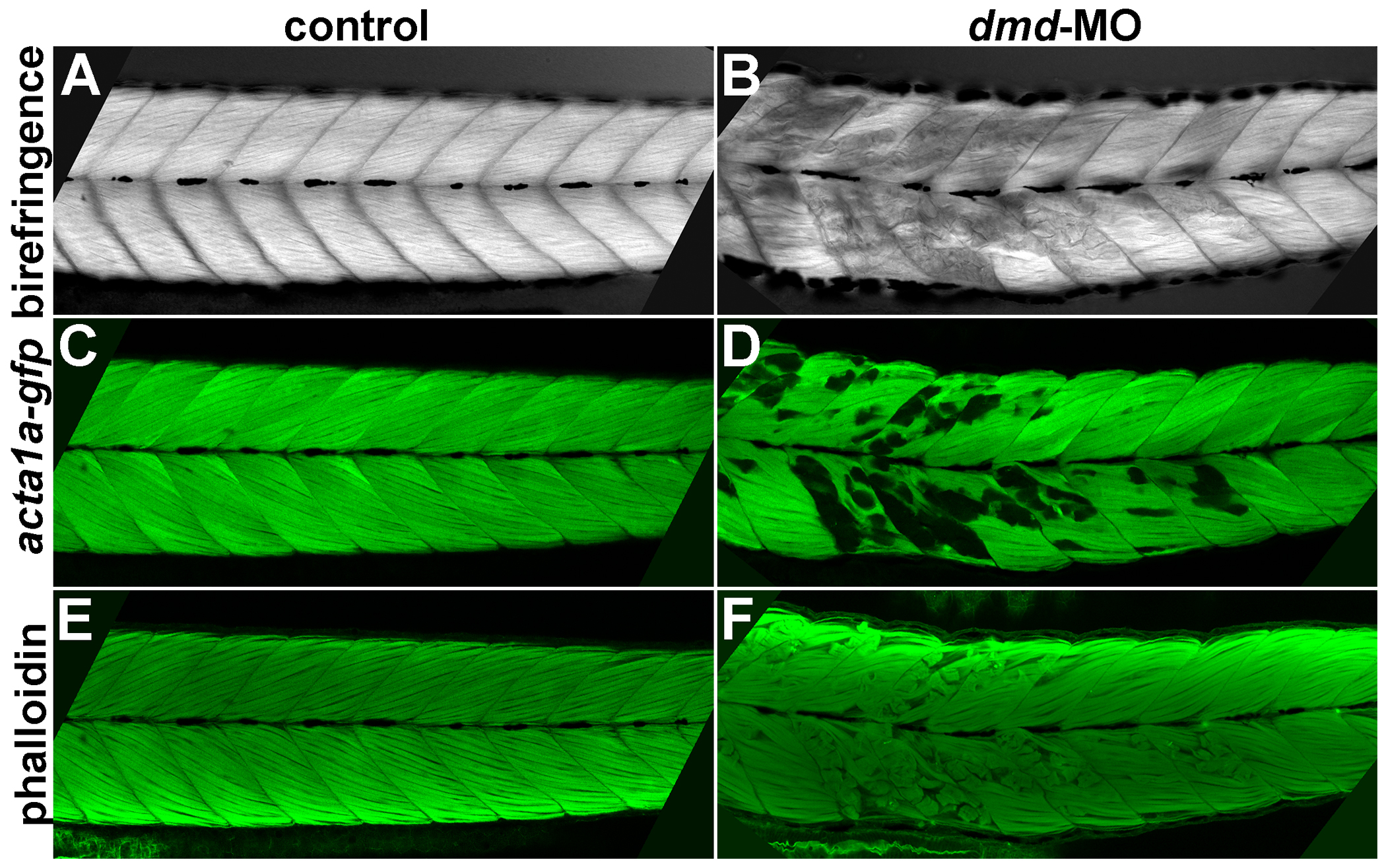
Correlation among birefringence, acta1a:gfp, and phalloidin patterns.
Confocal images of a single control (A,C,E) and dmd-MO (B,D,F) larva at 4 dpf. Phalloidin staining was imaged using the red channel but false-colored in green in E,F. Lateral views of trunk somites show anterior to the left. The birefringence, acta1a-gfp, and phalloidin muscle lesion patterns strongly correlate in all larvae that were examined for all three patterns (n=8). Abnormal birefringence also correlates with lesions visualized with phalloidin staining in dmd-/- larvae (n=13, not shown).
Figure Data
Acknowledgments
This image is the copyrighted work of the attributed author or publisher, and
ZFIN has permission only to display this image to its users.
Additional permissions should be obtained from the applicable author or publisher of the image.
Full text @ PLoS Curr.