
|
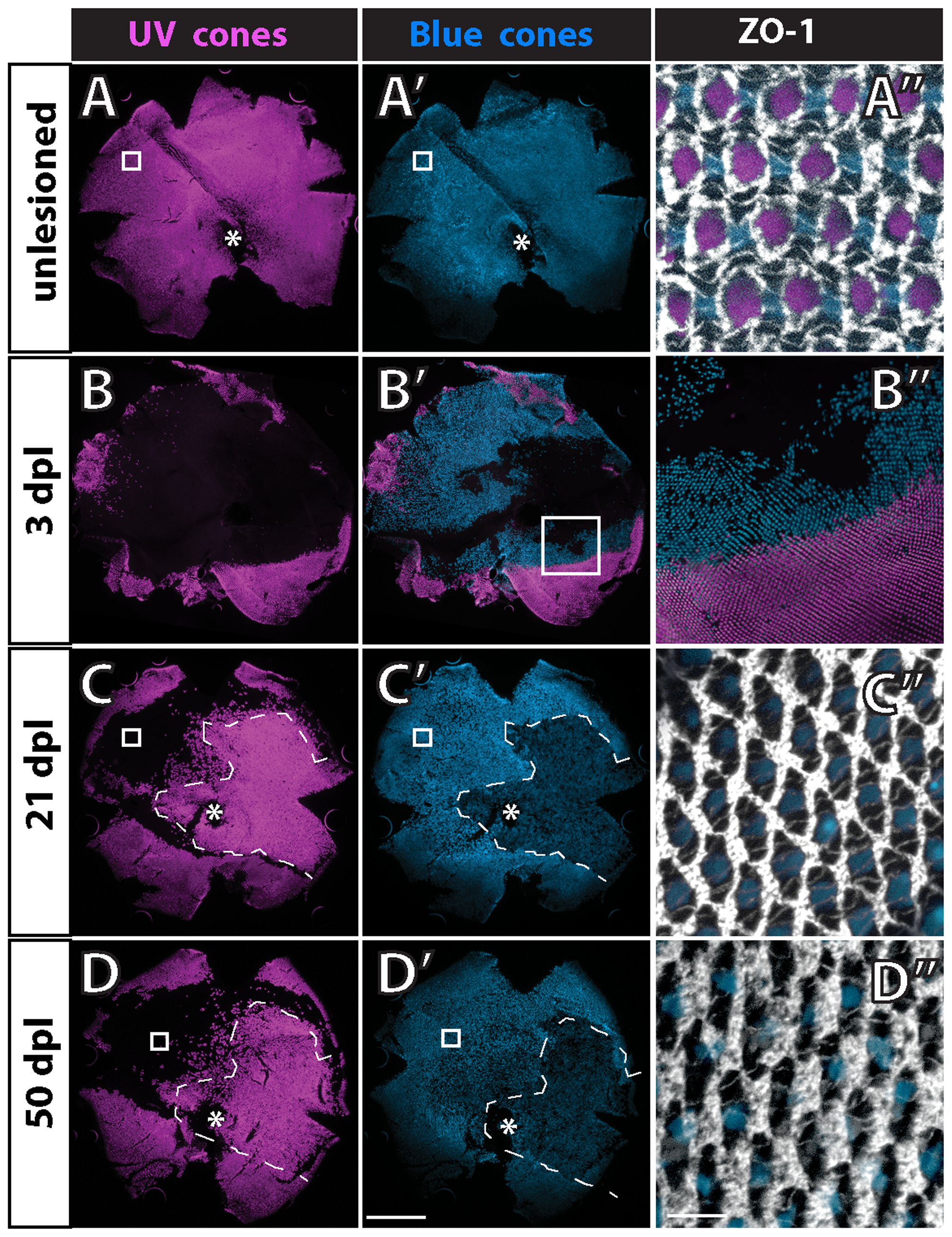
Fig. 10
The mosaic pattern is not disrupted after selective ablation of UV cones in adult wild-type fish.
Retinal flat-mounts from a double transgenic adult fish (UV and blue cone reporters) immunostained for ZO-1. Ventral is down; optic disc (*); boxed area in panels to the right. A) Control retina, UV cones; A2) blue cones; A3) ZO-1, UV and blue. B) Transgenic fish exposed to intense light, 3 days post-lesion (dpl), UV cones; B2) blue and UV cones; B3) boundary of ventral region in which no cones are ablated (lower right) and only UV cones but not blue cones are ablated (upper right). C) At 21 dpl, all cone types, including UV cones, were ablated and have regenerated within the central and temporal retina in the region enclosed by dashes; C2) blue cones have regenerated within the region enclosed by dashes; C3) in the boxed region where only UV cones were ablated they do not regenerate. D) At 50 dpl, UV cones (along with other cone types) have regenerated within the region enclosed by dashes; D2) blue cones have regenerated within the region enclosed by dashes; D3) rods continue to accumulate in the spaces previously occupied by UV cones in the region where they were selectively ablated and fail to regenerate. Scale bars: 500 μm (A, A2 through D, D2); 10 μm (A3 through D3).