
|
Fig. 1
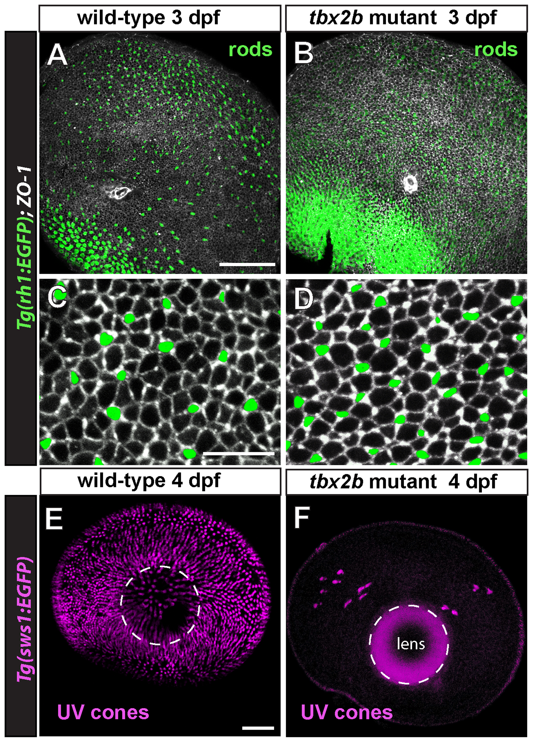
Rod photoreceptors develop rapidly and UV cones are missing in tbx2b mutants.
A) Isolated, larval rh1:EGFP eye at 3 days post-fertilization (dpf) viewed from the scleral aspect. Rod photoreceptors are green and immunostaining for the apical junctional marker, Zonula Occludens-1 (ZO-1) is in white. Dorsal is up; the optic nerve appears as a small white ring ventral to the center. B) Mutant tbx2b; rh1:EGFP larval eye at 3 dpf viewed from the scleral side. Note increased number of rods, especially in the ventral retina. C, D) Higher magnifications of central retina in wild-type and mutant eyes, respectively. E) Isolated, larval sws1:EGFP eye and F) tbx2b; sws1:EGFP eye at 4 dpf, viewed from the scleral aspect. Cones expressing the UV opsin reporter are pseudocolored magenta. The lens, outlined with a dashed white line, shows background fluorescence in F, due to the longer exposure time required to capture immunofluorescence of the few scattered UV cones. Scale bars: = 50 μm (A,B); 10 μm (C,D); 50 μm (E,F).