
|
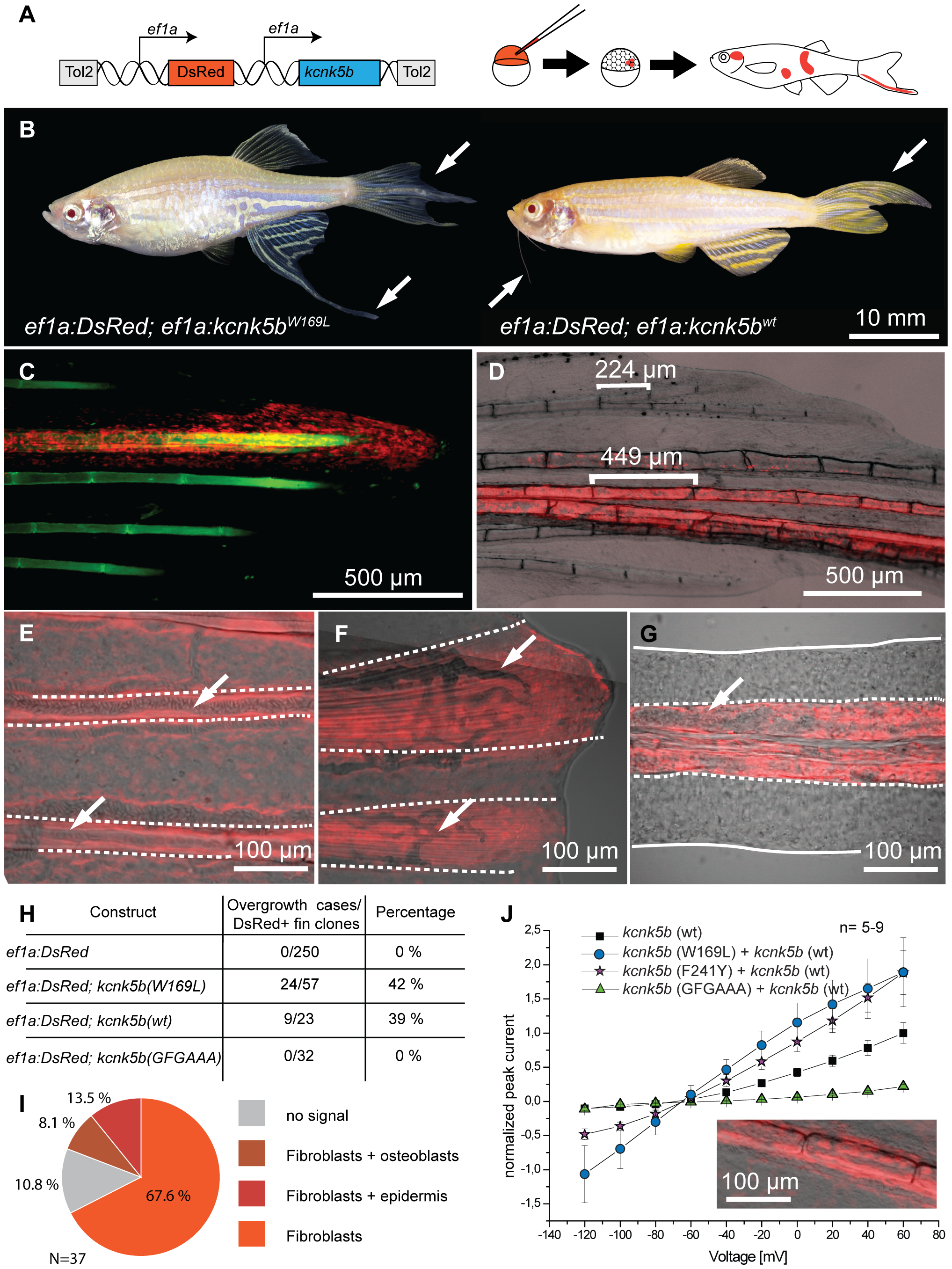
Fig. 6
Overexpression of kcnk5b is sufficient to cause fin overgrowth.
(A) Construct used to create kcnk5b-expressing clones via Tol2 transgenesis. (B) Individual fish expressing kcnk5b (W169L) (left) or kcnk5b (wt) (right) in mosaic clones display localized fin and barbel overgrowth. (C–F) Overgrowth is associated with DsRed expression (in red) within mesenchymal cells. (C) Calcein staining labels bone tissue (in green) of an overgrown fin (DsRed; kcnk5(W169L) expressing clone). (D) Mesenchymal clones are associated with increased segment length in the fin compared to non-overgrown DsRed negative regions. (E) Fibroblast-like cells appear as DsRed positive cells within the fin rays (dotted line) that surround DsRed negative vasculature (arrows in E and F) which extend along the actinotrichia (fibrils within dotted lines in F) towards the distal end of the fin. (G) Overgrown barbels show DsRed signal within the mesenchyme (area within dotted line) but not in the vasculature (arrow). (H) Number of clones associated with overgrowth in different kcnk5b variants. (I) Proportion of different cell types labeled in overgrown tissues. (J) Electrophysiological recordings of the non-conductive kcnk5b (GFGAAA) mutant in oocytes. Squares: kcnk5b (wt), purple stars: kcnk5b (F241Y)+kcnk5b (wt), blue circles: kcnk5b (W169L)+kcnk5b (wt), green triangles: + kcnk5b (GFGAAA)+kcnk5b (wt). Current was normalized to the measurement of wt current at 60 mV. Inset: DsRed+ fibroblasts in fish injected with the non-conductive construct do not lead to fin overgrowth.