
|
Fig. 6
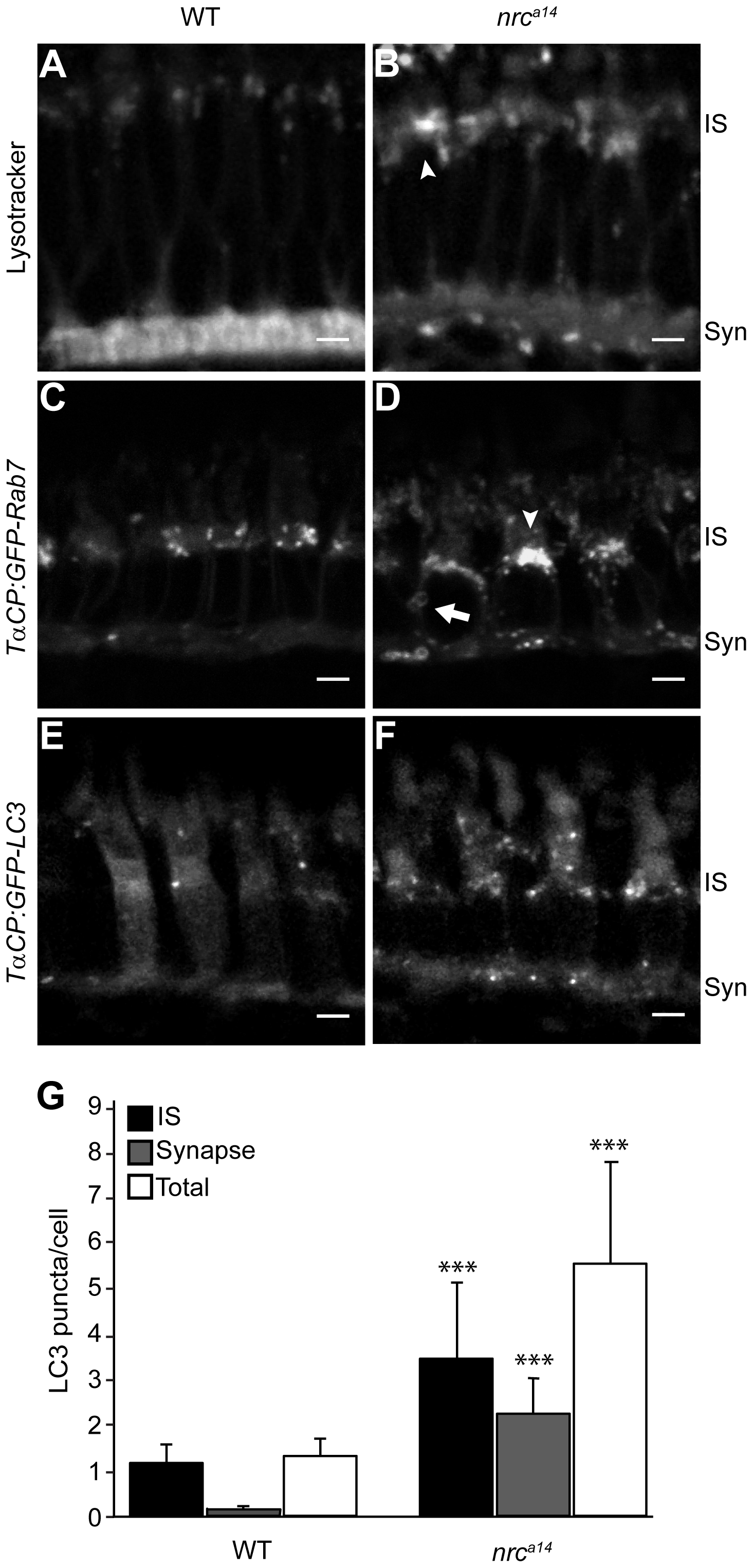
Loss of SynJ1 disrupts endolysosomal structures.
Confocal images of larvae incubated with Lysotracker Red and the transgenic fish lines Tg(TαCP:GFP-Rab7) and Tg(TαCP:GFP-LC3) mark the endolysosomal system. 5 dpf larvae were incubated in Lysotracker and imaged live. In WT retinas (A), Lysotracker Red accumulated primarily in the synapse and in small punctate structures in the IS of cone photoreceptors. In nrca14 cone photoreceptors (B), Lysotracker Red accumulated primarily in larger, abnormal structures in the IS. Retinal slices from WT (C) and nrca14 (D) larvae show that abnormal Rab7-positive structures including large perinuclear structures (arrowheads), and doughnut shaped structures (arrows) accumulated in the IS and synapse in the absence of SynJ1. The lack of SynJ1 also caused an increase in the number of LC3-GFP-positive structures, indicating a disruption in autophagy in nrca14 cones (F). The number of LC3 puncta is increased in both the IS and synapse of nrca14 cones (G). Syn = photoreceptor synapses, IS = inner segment. Scale bar = 2 μm in all images. Graph shows average LC3 puncta in the IS, synapse or entire cell (Total) per cell, error bars are STDEV for 11 WT larvae and 12 nrca14 larvae. The number of LC3 puncta per cell for each subcellular compartment or the entire cell was significantly different between WT and nrca14 larvae (p-value<0.001, donated by ***).