
|
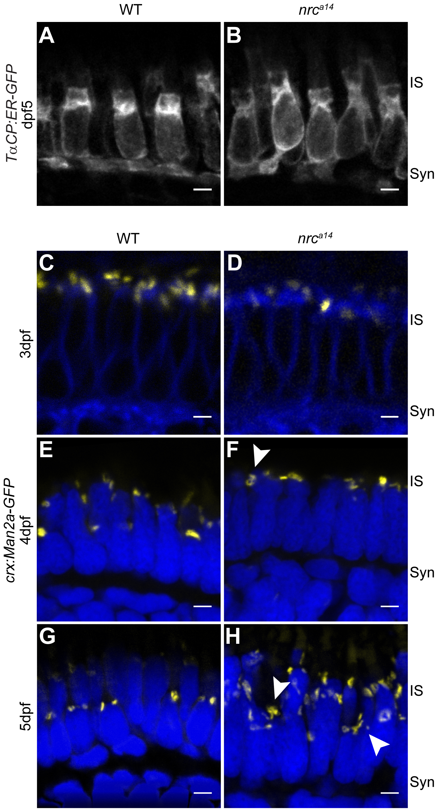
Fig. 5
SynJ1 is required for Golgi maintenance but not development.
The transgenic fish line Tg(TαCP:ER-GFP) was used to mark the ER. Retinal slices from 5 dpf WT (A) and nrca14 (B) larvae showed that the overall ER morphology was unaltered in the absence of SynJ1. Confocal images of Tg(crx:Man2a-GFP) WT (C, E, G) or nrca14 (D, F, H) larvae retina at 3–5 dpf showed that the Golgi is normal during photoreceptor development, but develops abnormalities disordered after cones become functional. No apparent abnormalities were seen in Golgi morphology of nrca14 compared to WT photoreceptors at 3 dpf (compare C and D). At 4 dpf, some mild morphology changes appeared in nrca14 Golgi (F, arrowhead). At 5 dpf, fragmented Golgi were visible in nrca14 photoreceptors (H, arrowheads). Man2a-GFP signal is shown in yellow, membranes of 3 dpf larvae were stained with BODIPY-TR and are shown in blue, and nuclei were stained with Hoechst and are shown in blue for 4 dpf and 5 dpf images. Syn = photoreceptor synapses, IS = inner segment. Scale bar = 2 μm in all images.