
|
Fig. 2
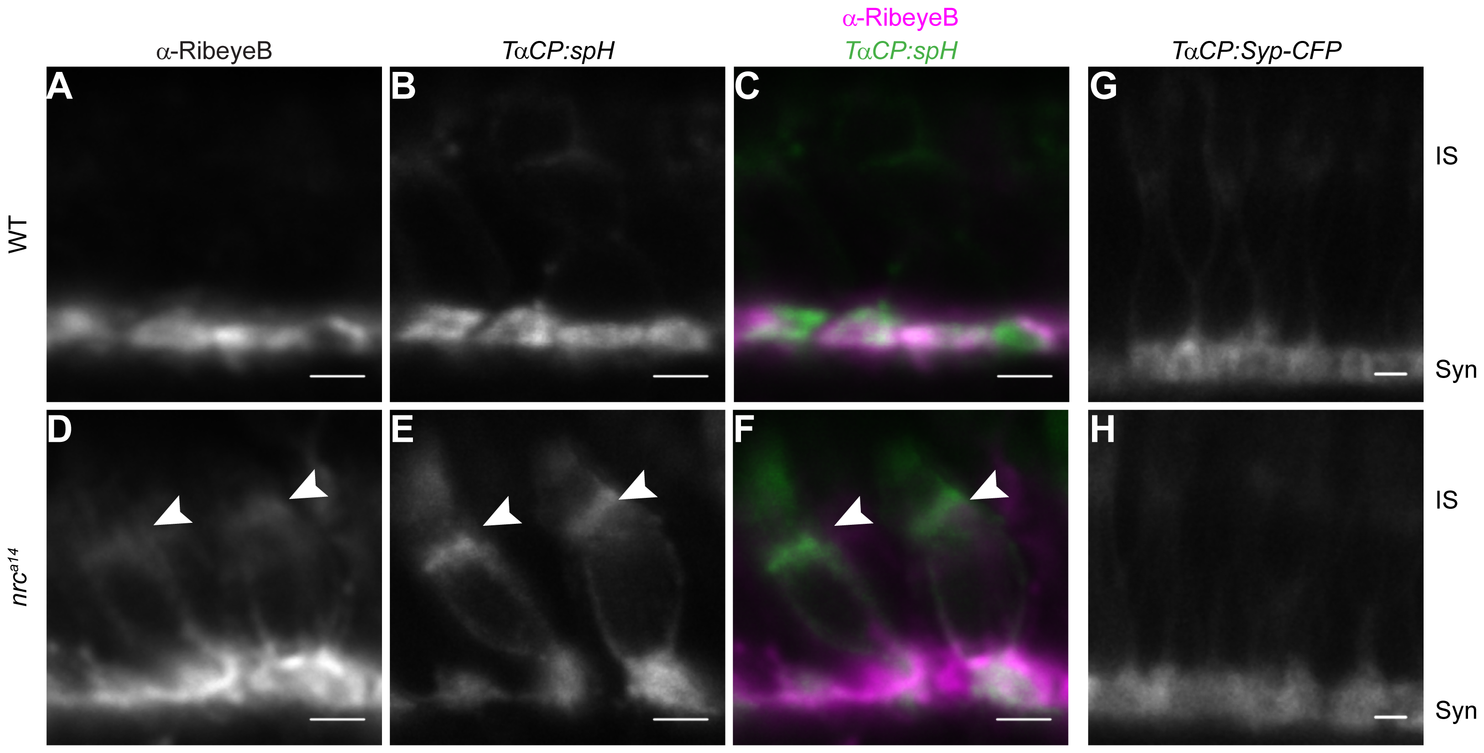
Some synaptic proteins are mislocalized in nrca14 cone photoreceptors.
Tg(TαCP:spH) WT and nrca14 5 dpf retinal slices were stained with an antibody against the ribbon synapse protein RibeyeB (A–F). In WT photoreceptors, RibeyeB was found only at synaptic terminals (A). In nrca14 cone photoreceptors, RibeyeB (D, arrowhead) and VAMP2 (E, arrowhead) were detected in both synaptic terminals and ISs. Mislocalized signals for RibeyeB (magenta) and VAMP2 (green) were coincident in the ISs of nrca14 cone photoreceptors (F, arrowhead). In contrast, the synaptic vesicle protein Synaptophysin (Syp-CFP) had a primarily synaptic distribution in both WT (G) and nrca14 (H) cone photoreceptors. Live confocal images were taken of 5 dpf Tg(TαCP:Syp-CFP) WT and nrca14 larvae. Syn = photoreceptor synapses, IS = inner segment. Scale bar = 2 μm.