
|
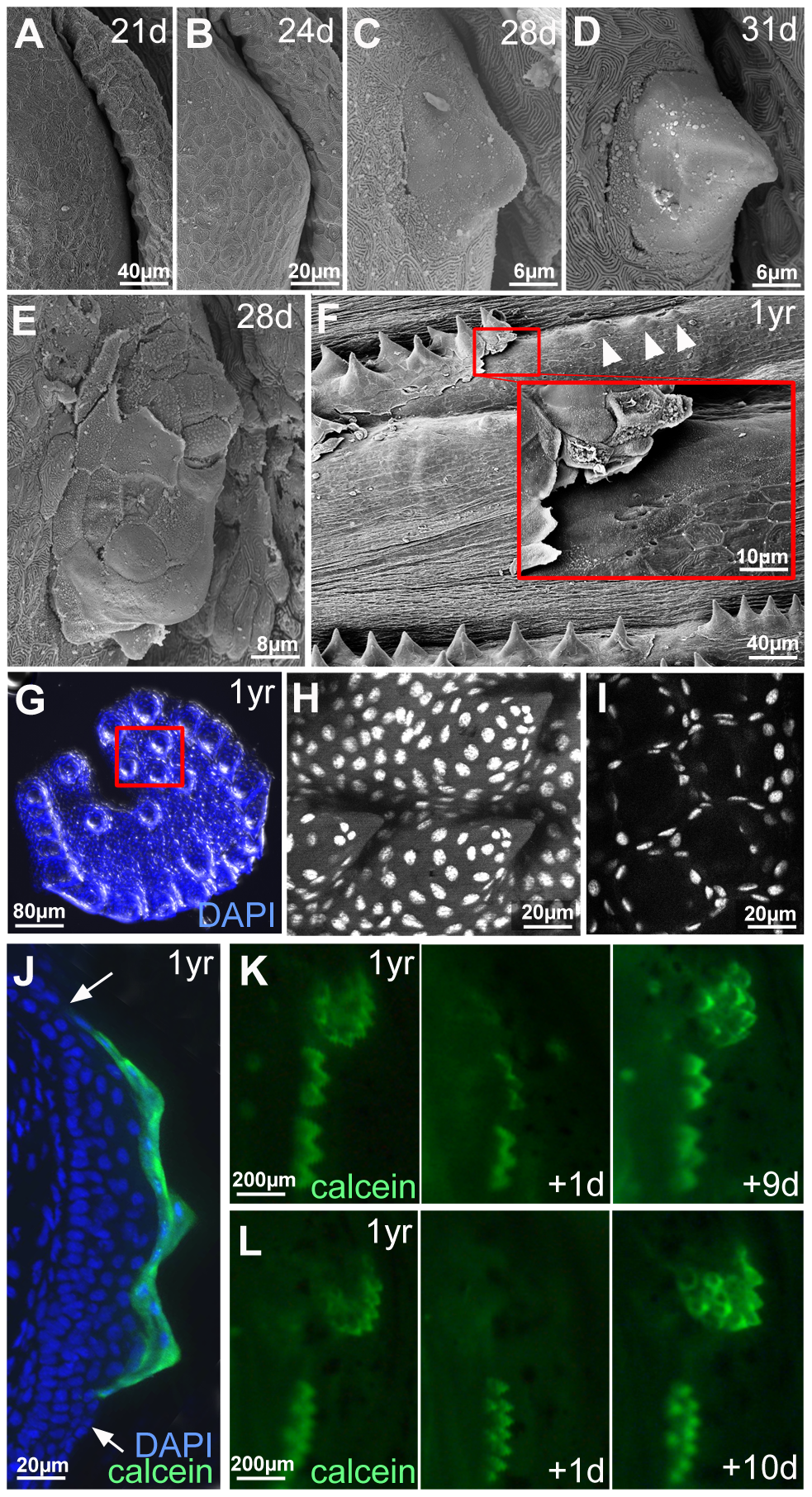
Fig. 4
Outer cells of breeding tubercles undergo desquamation and renewal.
(A–D) SEM images showing developmental time course of breeding tubercle formation on lower jaw between 21 and 31 days of development. (E) SEM image of a breeding tubercle on the lower jaw at 28 days of development. In the upper part of the image superficial cells have been lost, and former second tier cells are exposed. (F) SEM image of breeding tubercles on the pectoral fin of a male fish; 1 year of age. Inset shows magnification of boxed region. Entire rows of breeding tubercle cap layers are lifted up. Directly after desquamation of the cap layer, tubercles have a more shallow and non-spiky shape (arrowheads). (G–I) Shed cap layers of tubercles of disc-like structure on lower jaw, found in the water after spawning of 1 year old fish; stained with DAPI. Region boxed in (G) is shown in (H–I) as confocal micrographs at higher magnification; (H) maximal projection; (I) single plane. (J) Transverse cryosection through the breeding tubercles of the lower jaw of a 1 year old fish that had been incubated in calcein solution; calcein fluorescence in green; nuclei stained with DAPI in blue. Arrows point to outer layer of regular epidermis. (K,L) Calcein fluorescence of breeding tubercle region on the lower jaw of live fish, 1 year of age, repetitively stained with calcein and monitored daily. Days after first image acquisition are indicated (+1 d, +9 d, +10 d).