
|
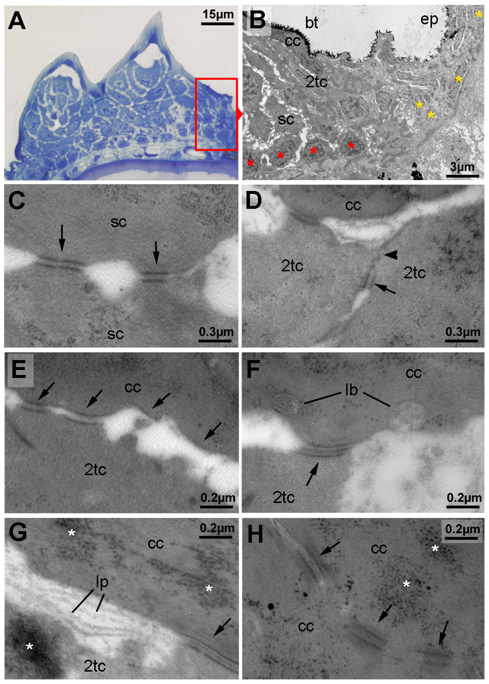
Fig. 3
Ultrastructure of different tubercle cell layers.
(A) Semi-thin section through breeding tubercles of lower jaw of a 1 year old wild-type, stained with methylene blue. For schematic overview, see also Figure 9A. Parts of the region shown in (B) are framed in red. (B–H) Transmission electron micrograph of ultrathin sections of same specimen as in (A). (B) Marginal zone of breeding tubercle. In the breeding tubercle (bt), basal cells are more regularly aligned (red asterisks) than in the adjacent epidermis (ep; yellow asterisks). Spinous (sc), second-tier (2tc) and outer cap cells (cc) are indicated. (C) Interphase between two spinous cells. Arrows point to desmosomes. (D) Interphase between two second-tier cells. At their apical side, cells are sealed to each other via tight junctions (arrowhead), directly followed by a desmosome (arrow), displaying the same spatial organization as in peridermal cells of regular epidermis (not shown) [12]. (E–G) Interphase between second-tier and outer cap cell, displaying progressive desmosomal regression (arrows) (E), extrusion of lamellar-body-like vesicles (lb) into the extracellular space (F), which is filled with material reminiscent of lipid lamellae (lp; G). Arrows point to desmosomes, asterisks mark highly abundant protein aggregates in cap and second tier cells (also in H). (H) Interphase between two outer cap cells, displaying progressive desmosomal regression and cell membrane deterioration. Abbreviations: 2tc, second-tier cell; cc, cap cell; lb, lamellar body; lp, lipid lamella; sc, spinous cell.