
Fig. 5
- ID
- ZDB-IMAGE-140730-91
- Genes
- Publication
- Kapp et al., 2013 - The integrator complex subunit 6 (ints6) confines the dorsal organizer in vertebrate embryogenesis
- All Figures
- Figures for Kapp et al., 2013
|
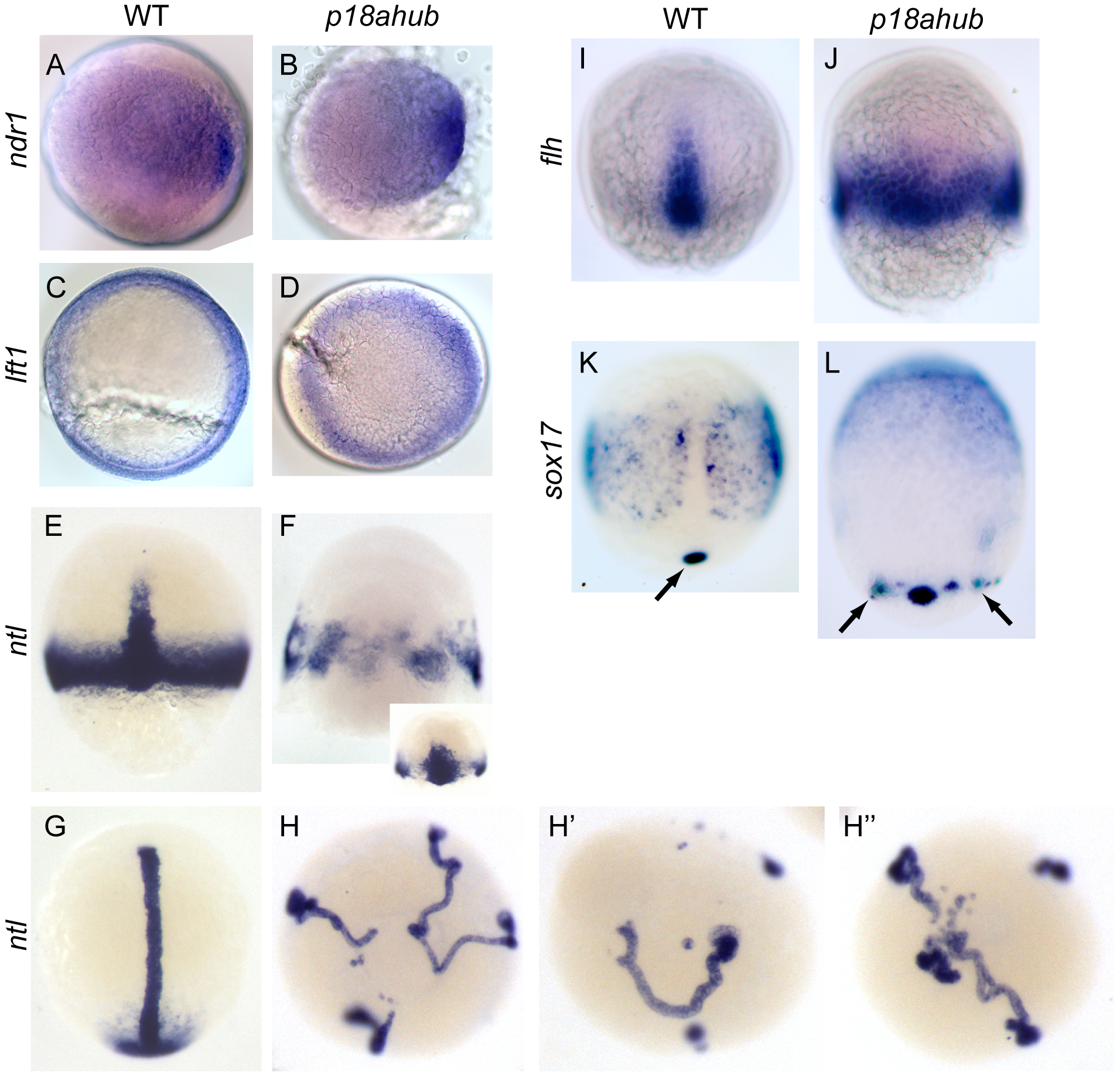
Fig. 5
Excessive axial mesoderm at the expense of ventrolateral mesoderm in p18ahub embryos.
WT (A, n = 19) and stage-matched p18ahub (B, n = 20) embryos at a mid blastula stage, exhibited similar nodal related 1 (ndr1) expression. WT (C, n = 14) and age-matched p18ahub (D, n = 13) embryos at 6 hpf after synchronized matings displayed similar lefty1 (lft1) expression, although blastoderm involution was not evident in the mutants. (A–D) are animal pole views, dorsal to right. (E–L) are mid gastrula stage, except (G and H) are 3–5 somite stage or equivalent. Dorsal views, animal to top, except (H–H3) are animal pole views. WT embryos (E, n = 6) displayed marginal and axial ntl expression. In contrast, some p18ahub embryos displayed only axial ntl expression distributed circumferentially (F, n = 16), while others displayed broadened axial and reduced ventrolateral ntl expression (F inset, n = 8). In WT embryos (G, n = 22) ntl was expressed in the notochord and tail bud mesenchyme. In equivalent stage p18ahub embryos, (H–H3, n = 21), ntl expression was observed in multiple notochords terminating in smaller tail bud-like domains. Consistent with ectopic axial ntl expression, the expression of floating head (flh), a marker of notochord precursors, confined dorsally in WT embryos (I, n = 20), was expressed around the entire embryonic margin in p18ahub embryos (J, n = 16). In WT (K, n = 18) sox17 is expressed in endodermal precursor cells and in a single cluster of dorsal forerunner cells (arrow). In p18ahub embryos (L, n = 16) sox17 expression indicates the presence of ectopic clusters of dorsal forerunner cells (arrows).