
Fig. 3
- ID
- ZDB-IMAGE-140730-89
- Genes
- Antibodies
- Publication
- Kapp et al., 2013 - The integrator complex subunit 6 (ints6) confines the dorsal organizer in vertebrate embryogenesis
- All Figures
- Figures for Kapp et al., 2013
|
Fig. 3
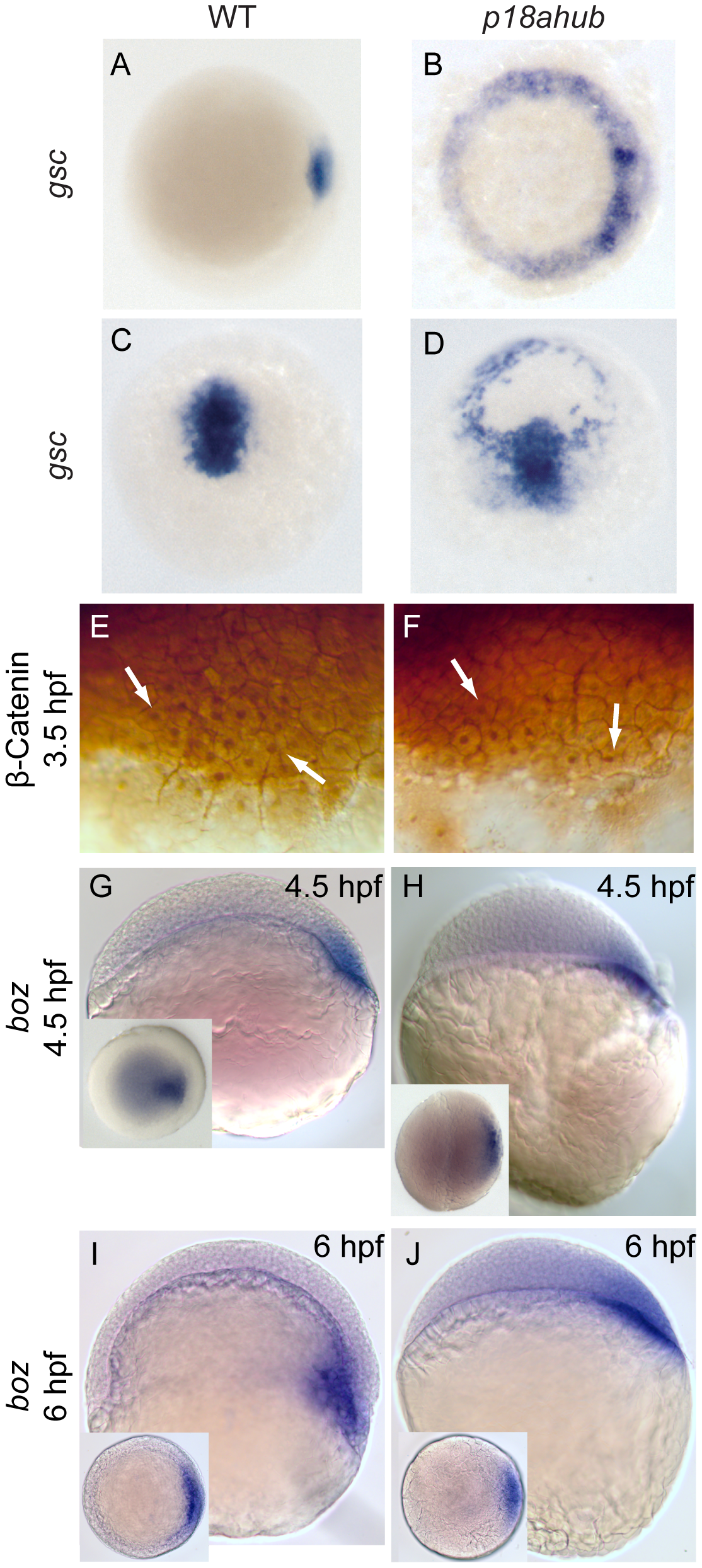
Dorsal organizer gene expression is expanded in p18ahub mutants
In situ hybridization using goosecoid (gsc) probe (A–D). Animal pole views, dorsal to right for (A) and (B); dorsal view, animal pole up in (C) and (D). WT embryos at early gastrula (A, n = 27) and mid gastrula stages (C, n = 41). p18ahub embryos (B, n = 33 and D, n = 10) displayed radially expanded gsc expression at the same stages. The embryo in (D) is tilted toward the viewer to show radial gsc expression. WT (E, n = 18) and p18ahub (F, n = 18) blastula stage embryos showed no obvious differences in the number or distribution of β-catenin immunopositive nuclei (arrows). (G–J) In situ hybridization for bozozok (boz), lateral views, dorsal to right; insets show animal pole views. WT and p18ahub embryos showed normal boz expression at late blastula (G, n = 19 and H, n = 18) and early gastrula stages (I, n = 14 and J, n = 16, respectively). As described in the text, epiboly progression was delayed in p18ahub embryos. At 6 hpf WT embryos form a morphologically apparent organizer, indicated by a thickening of the blastoderm on the dorsal side of the embryo and boz expression within the hypoblast. In contrast, at 6 hpf p18ahub embryos still appeared to be in a late blastula stage, although their boz expression was similar to WT.