
Fig. 1
- ID
- ZDB-IMAGE-140730-87
- Publication
- Kapp et al., 2013 - The integrator complex subunit 6 (ints6) confines the dorsal organizer in vertebrate embryogenesis
- All Figures
- Figures for Kapp et al., 2013
|
Fig. 1
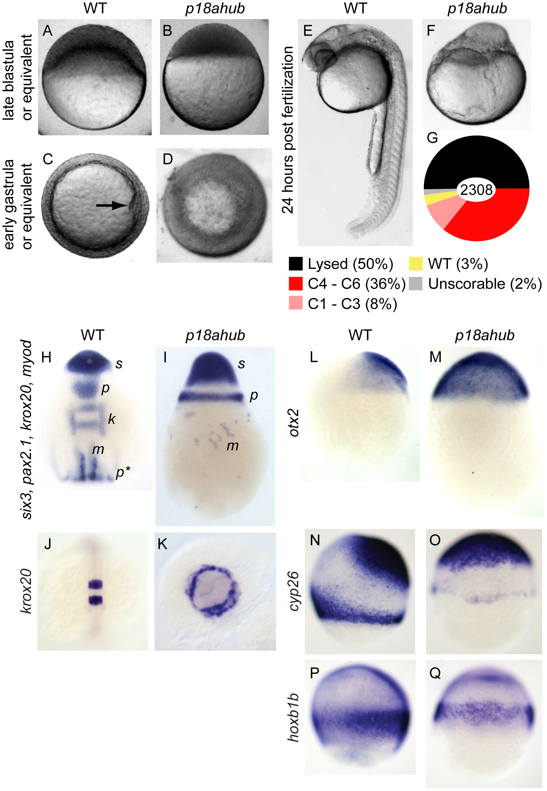
p18ahub mutants exhibit delayed progression of epiboly and severe dorsalization.
Late blastula stage WT embryo (A), and age-matched p18ahub embryo (B). WT embryo at an early gastrula stage (C) with the shield (organizer) on the dorsal side (arrow). A p18ahub embryo at an early gastrula stage (D) displaying apparent radial hyperconvergence with no definitive dorsal side. (A,B) lateral views; (C,D) animal pole views. WT (E) and p18ahub (F) embryos at 24 hours post fertilization (hpf) (lateral views). (G) Pie chart depicting proportion of embryos with indicated phenotypes at 24 hpf sampled over a total of 2308 embryos. (H and I), in situ hybridization on 3–5-somite stage WT (H, n = 15) and age-matched p18ahub (I, n = 13) embryos for six3 (s) expression in presumptive forebrain, pax2.1 expression in the midbrain-hindbrain boundary (p) and pronephros (p*), krox20 (k) in hindbrain rhombomeres 3 and 5, and myod (m) in paraxial mesoderm; dorsal view in (H), lateral view in (I), anterior to top. (J and K), 10-somite stage WT (J, n = 17, dorsal view), and age-matched p18ahub embryos (K, n = 18, anterior view) processed for krox20 in situ hybridization. (L–Q) in situ hybridization on mid gastrula stage WT and equivalent stage p18ahub embryos shown for: otx2, WT (L, n = 18) and p18ahub (M, n = 10); cyp26a, WT (N, n = 16) and p18ahub (O, n = 15); and hoxb1b, WT (P, n = 22) and p18ahub (Q, n = 21). Lateral views, anterior at top, dorsal to right.