
|
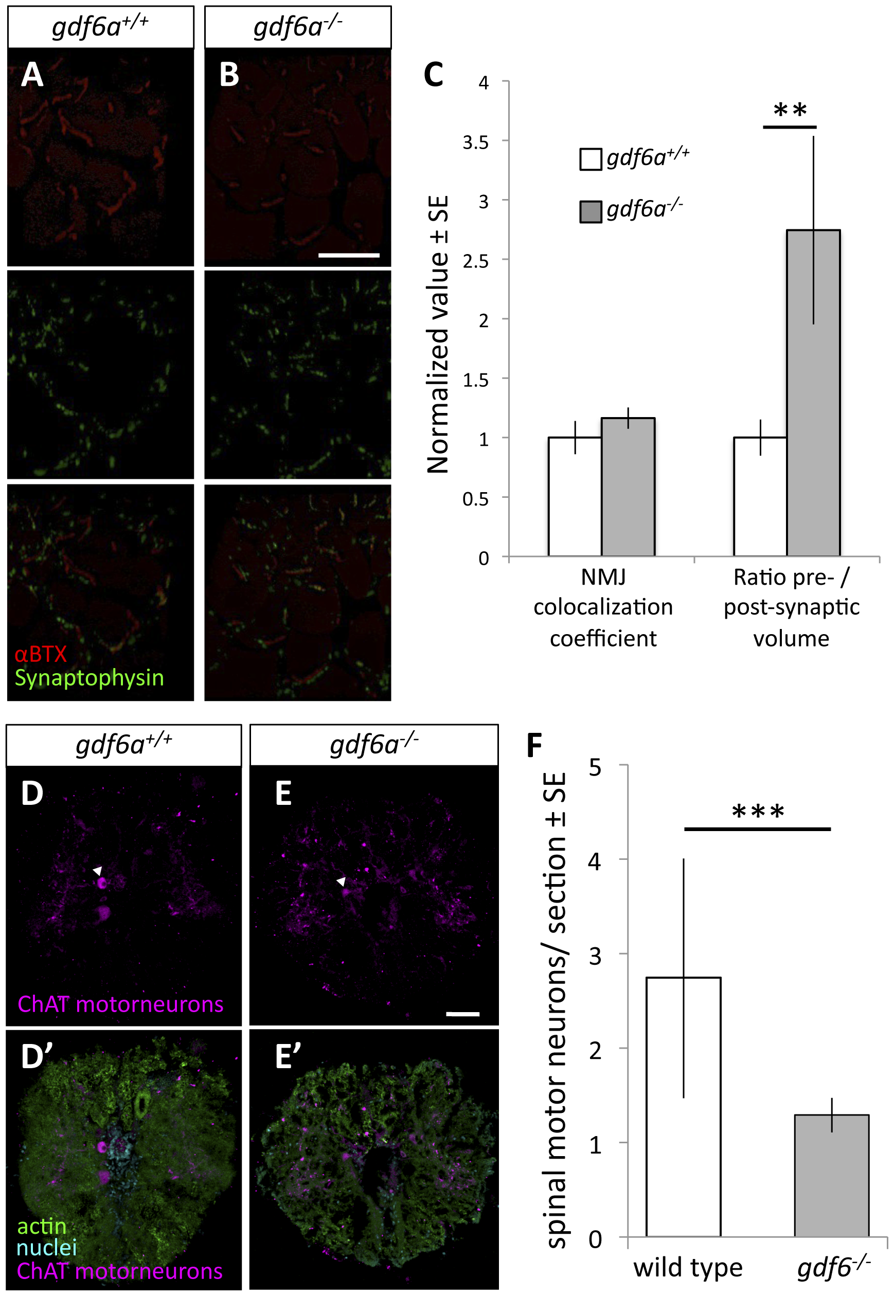
Fig. 3
Zebrafish harboring homozygous mutations in gdf6 exhibit altered neuromuscular junctions and fewer spinal motor neurons.
A-B. Assessment of neuromuscular junction (NMJ) morphology including presynaptic (synaptophysin) and postsynaptic (αBTX) compartments in 9 month old gdf6-/- and gdf6+/+ siblings by immunohistochemistry. C. ALS-like increases in motoneuron pre-synaptic volumes are observed in gdf6-/- fish when normalized to post-synaptic volumes (**p = 0.009, n = 5 fish per genotype). Coefficients of colocalization for pre- and post-synaptic compartments are not altered, as is expected in later-stage zebrafish ALS models. D-E. Motor neuron cell bodies were identified in cross-sections of spinal cord using immunohistochemistry against choline acetyl transferase (ChAT, e.g. arrowheads) in nine month old gdf6+/+ and gdf6-/- fish (panels A and B, respectively). Bottom panels affirm motoneuron cell body identification using actin and nuclear counter-stains. F. gdf6-/- fish have approximately 50% the abundance of spinal motor neurons compared to sibling gdf6+/+ fish. (Mann-Whitney U Test, *** p<0.001, n>50 sections from 4 fish per genotype, researcher blinded to genotype during quantification). Scale bar = 60 μm in A,B and 50 μm in D,E.