
Image
|
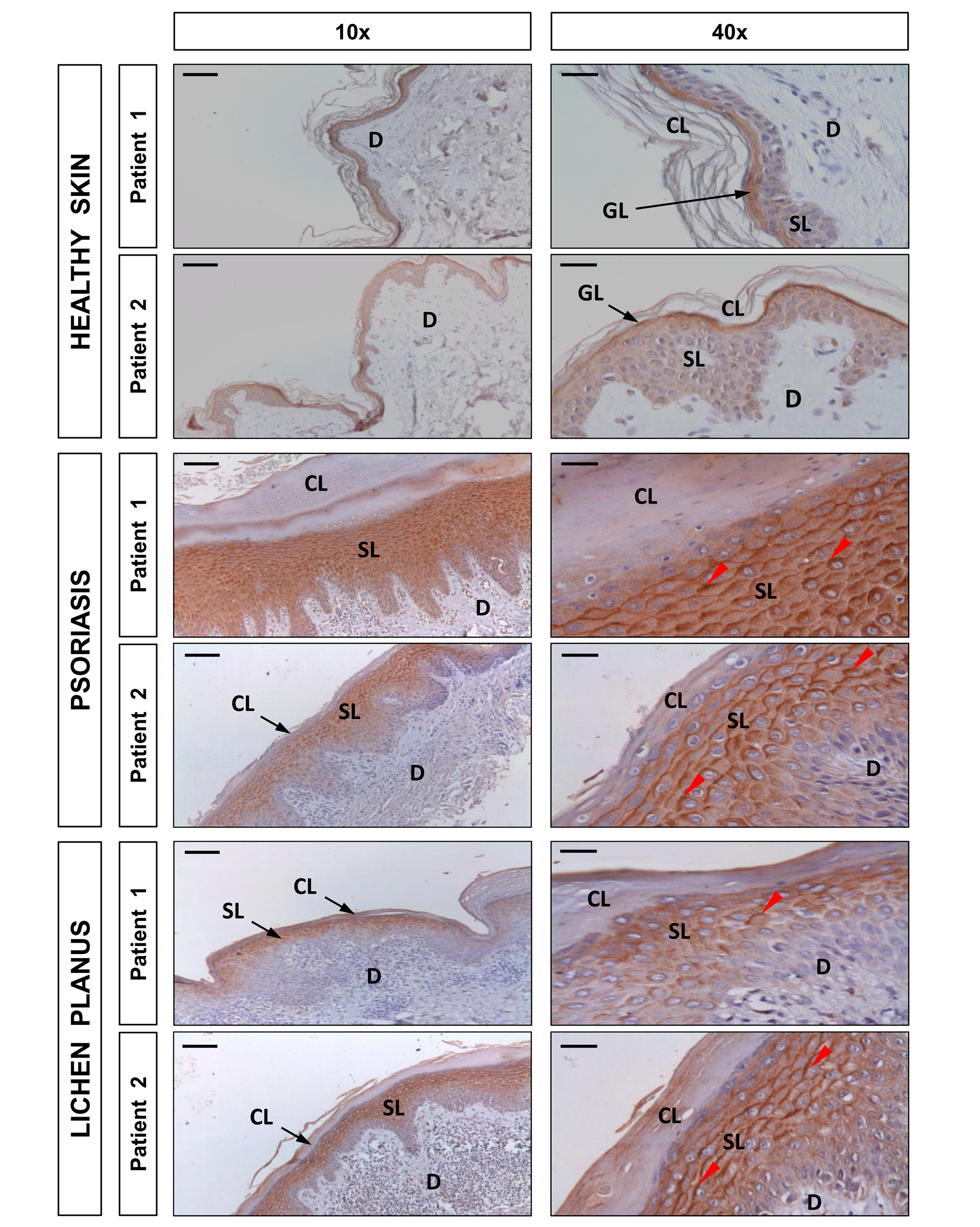
Figure Caption
Fig. 7
DUOX1 is induced in human psoriasis and lichen planus lesions.
Representative images of sections from two healthy, two psoriatic, and two lichen planus skin biopsies that have been immunostained with an anti-DUOX1 goat polyclonal antibody and then slightly counterstained with hematoxilin. Note that DUOX1 is weakly expressed in healthy epidermis, mainly in the granular layer (GL), whereas it is strongly expressed (red arrowheads) in the spinous layer (SL) of both psoriasis and lichen planus lesions. CL, cornified layer; D, dermis. Scale bars, 100 μm (left panel) and 30 μm (right panel).
Acknowledgments
This image is the copyrighted work of the attributed author or publisher, and
ZFIN has permission only to display this image to its users.
Additional permissions should be obtained from the applicable author or publisher of the image.
Full text @ PLoS Biol.