
Image
|
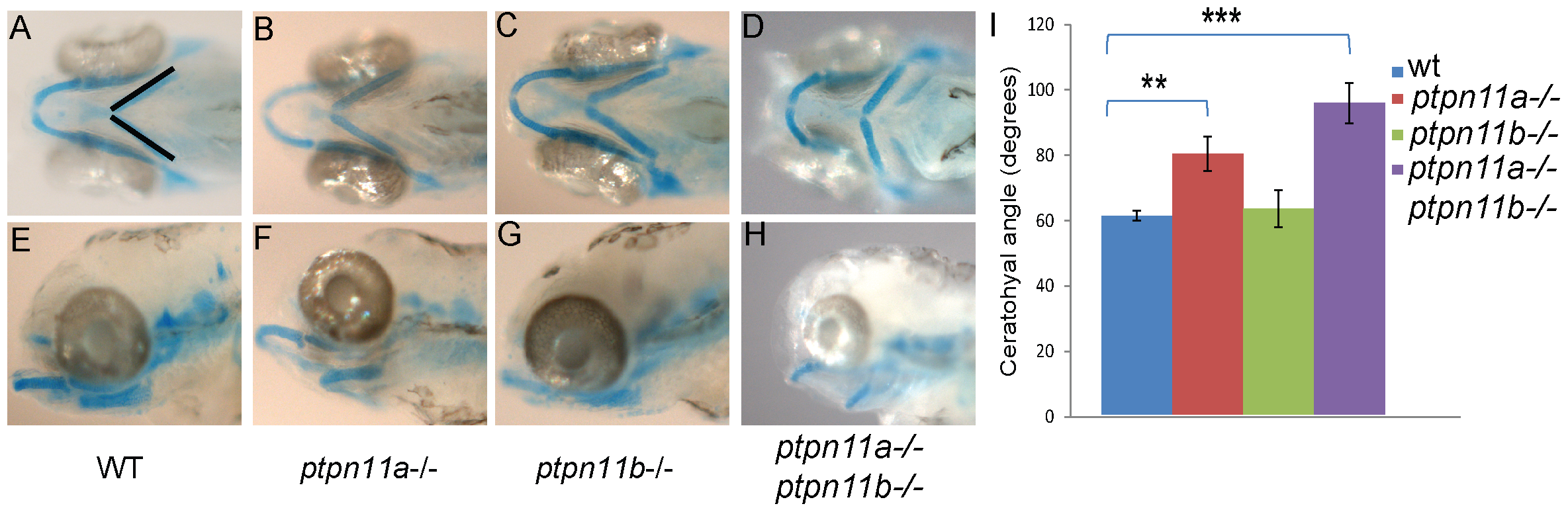
Figure Caption
Fig. 5
Ptpn11a-/- and double mutant embryos exhibit craniofacial defects at 5 dpf.
Alcian blue staining was done to visualize the cartilage. (a,e) wild-type, (b,f) ptpn11a-/-, (c,g) ptpn11b-/- and (d,h) double homozygous mutant. (i) Average width of the ceratohyal angle (indicated in panel a) was determined for each genotype (n = 10–24). Statistics were determined using a student′s t-test; ** indicates a p value of <0.01, *** indicates a p value <0.001.
Figure Data
Acknowledgments
This image is the copyrighted work of the attributed author or publisher, and
ZFIN has permission only to display this image to its users.
Additional permissions should be obtained from the applicable author or publisher of the image.
Full text @ PLoS One