
IMAGE
Fig. S3
- ID
- ZDB-IMAGE-140728-18
- Publication
- Acosta et al., 2014 - Mutant Human FUS Is Ubiquitously Mislocalized and Generates Persistent Stress Granules in Primary Cultured Transgenic Zebrafish Cells
- All Figures
- Figures for Acosta et al., 2014
Image
|
Figure Caption
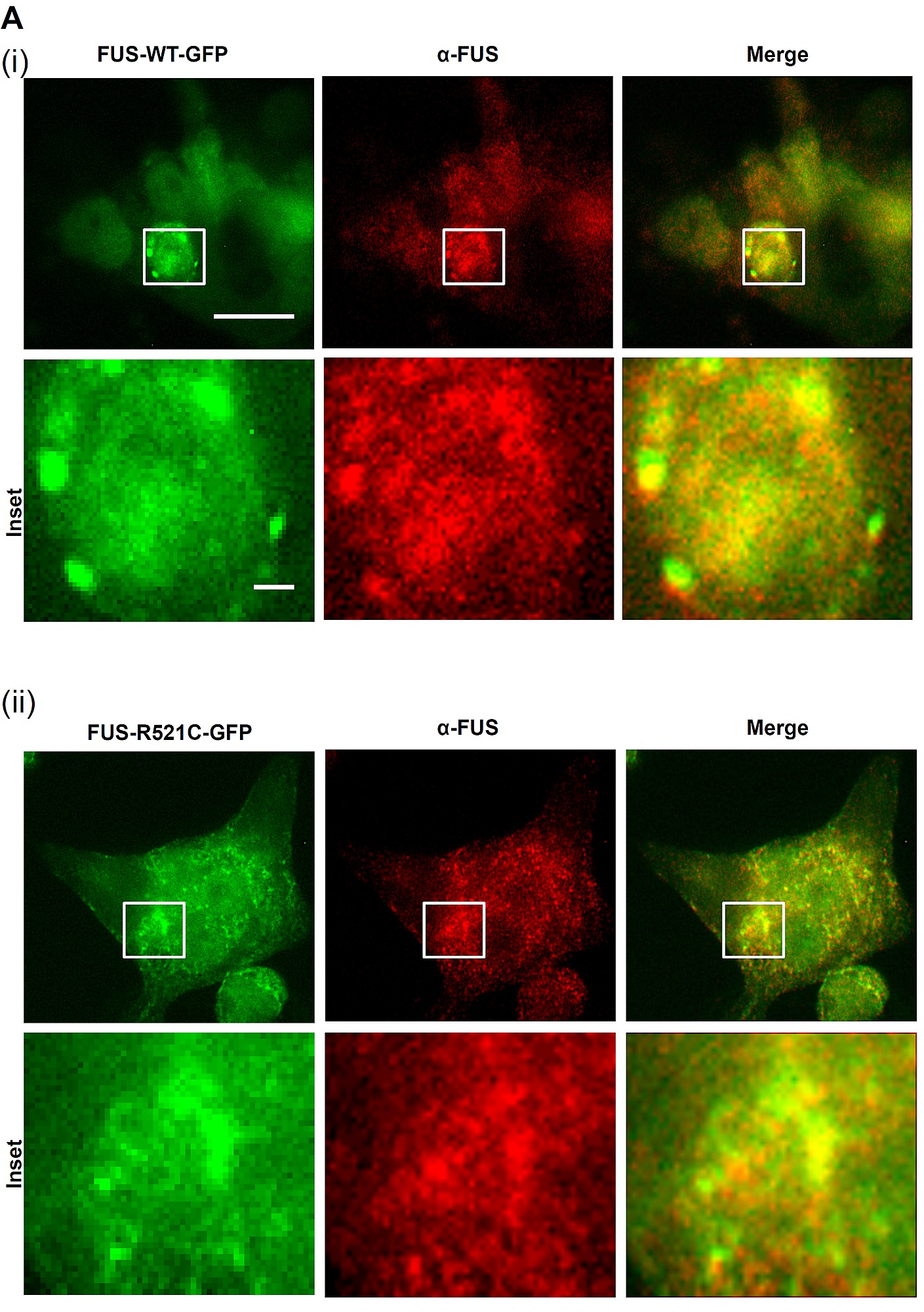
Fig. S3
Co-localization of FUS-GFP SGs and FUS (stained with polyclonal anti-FUS antibody, red) confirms the presence of human FUS in SGs. Scale bar = 20 μm; insets = 1 μm. Brand M, Heisenberg CP, et al. (1996) Mutations in zebrafish genes affecting the formation of the boundary between midbrain and hindbrain. Development 1236179–190.
Acknowledgments
This image is the copyrighted work of the attributed author or publisher, and
ZFIN has permission only to display this image to its users.
Additional permissions should be obtained from the applicable author or publisher of the image.
Full text @ PLoS One