
Fig. 1
- ID
- ZDB-IMAGE-140728-12
- Genes
- Publication
- Acosta et al., 2014 - Mutant Human FUS Is Ubiquitously Mislocalized and Generates Persistent Stress Granules in Primary Cultured Transgenic Zebrafish Cells
- All Figures
- Figures for Acosta et al., 2014
|
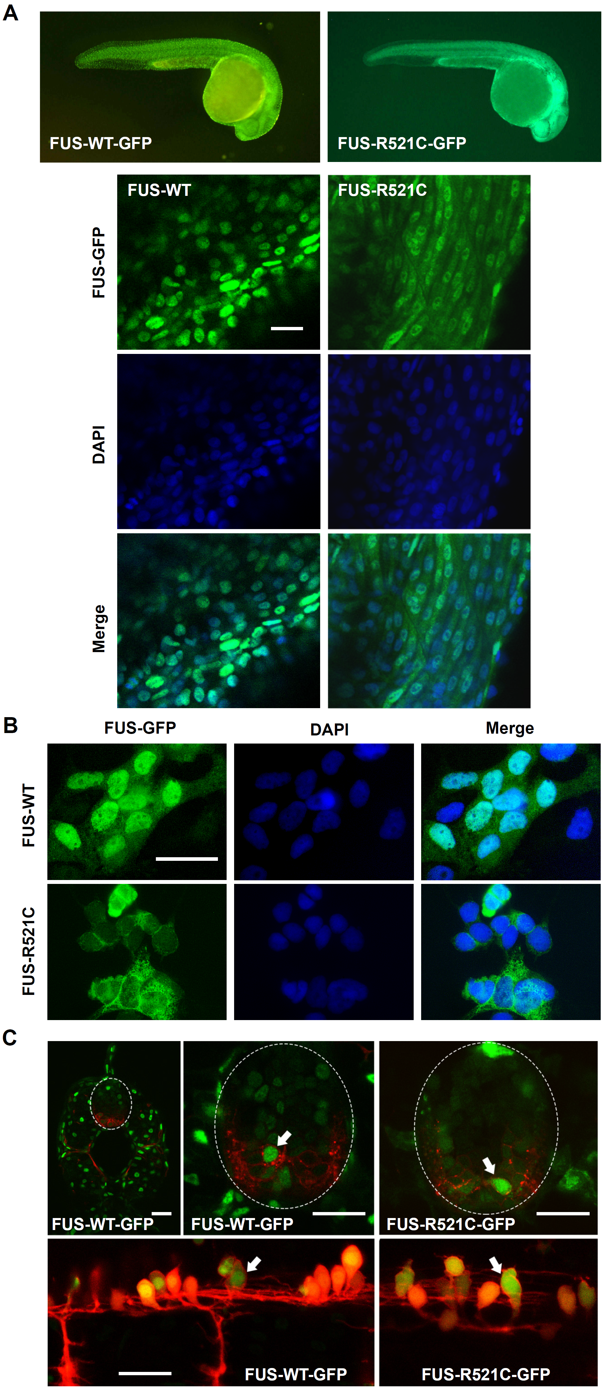
Fig. 1
Whole mount and cell cultures of FUS-GFP transgenic zebrafish.
(A) Transgenic zebrafish larvae whole mounts showed cytosolic mislocalization of mutant human FUS in FUS-R521C-GFP in comparison to FUS-WT-GFP which was restricted to cell nuclei. (B) FUS-R521C-GFP showed greater cytosolic distribution in comparison to FUS-WT-GFP in zebrafish primary cell cultures. (C) Confocal images of 48 hpf transgenic zebrafish spinal cord further demonstrate mislocalization of mutant FUS-521C-GFP (green) in motor neurons (red) (arrows). Images are maximum projections captured using a Leica SPE5 confocal microscope. Sagittal sections (upper images) of Tg(s1020tGAL4: UASmCherry) (Scott and Baier, 2009) and transverse sections (lower images) of Tg(HB9: mK02caax) membrane localised mk02 expressed in motorneurons by HB9 promoter (Flanagan-Steet et al 2005) with either FUS-WT-GFP or FUS-521C-GFP as indicated. Scale bar = 20 μm.